1L5B | pdb_00001l5b

DOMAIN-SWAPPED CYANOVIRIN-N DIMER


Experimental Data Snapshot

  • Method: X-RAY DIFFRACTION
  • Resolution: 2.00 Å
  • R-Value Free: 
    0.258 (Depositor), 0.259 (DCC) 
  • R-Value Work: 
    0.247 (Depositor), 0.246 (DCC) 

Starting Model: experimental
View more details

wwPDB Validation 3D Report Full Report

Validation slider image for 1L5B

This is version 1.4 of the entry. See complete history

Literature

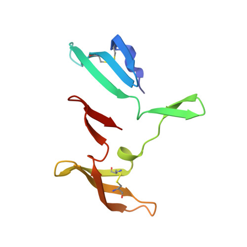
The domain-swapped dimer of cyanovirin-N is in a metastable folded state: reconciliation of X-ray and NMR structures.

Barrientos, L.G.Louis, J.M.Botos, I.Mori, T.Han, Z.O'Keefe, B.R.Boyd, M.R.Wlodawer, A.Gronenborn, A.M.

(2002) Structure 10: 673-686

  • DOI: https://doi.org/10.1016/s0969-2126(02)00758-x
  • Primary Citation Related Structures: 
    1L5B, 1L5E

  • PubMed Abstract: 

    The structure of the potent HIV-inactivating protein cyanovirin-N was previously found by NMR to be a monomer in solution and a domain-swapped dimer by X-ray crystallography. Here we demonstrate that, in solution, CV-N can exist both in monomeric and in domain-swapped dimeric form. The dimer is a metastable, kinetically trapped structure at neutral pH and room temperature. Based on orientational NMR constraints, we show that the domain-swapped solution dimer is similar to structures in two different crystal forms, exhibiting solely a small reorientation around the hinge region. Mutation of the single proline residue in the hinge to glycine significantly stabilizes the protein in both its monomeric and dimeric forms. By contrast, mutation of the neighboring serine to proline results in an exclusively dimeric protein, caused by a drastic destabilization of the monomer.


  • Organizational Affiliation
    • Laboratory of Chemical Physics, National Institute of Diabetes and Digestive and Kidney Diseases, National Institutes of Health, Bethesda, MD 20892, USA.

Macromolecule Content 

  • Total Structure Weight: 22.27 kDa 
  • Atom Count: 1,749 
  • Modeled Residue Count: 202 
  • Deposited Residue Count: 202 
  • Unique protein chains: 1

Macromolecules

Find similar proteins by:|  3D Structure
Entity ID: 1
MoleculeChains  Sequence LengthOrganismDetailsImage
cyanovirin-N
A, B
101Nostoc ellipsosporumMutation(s): 0 
UniProt
Find proteins for P81180 (Nostoc ellipsosporum)
Explore P81180 
Go to UniProtKB:  P81180
Entity Groups
Sequence Clusters30% Identity50% Identity70% Identity90% Identity95% Identity100% Identity
UniProt GroupP81180
Sequence Annotations
Expand
Reference Sequence

Small Molecules

Experimental Data & Validation

Experimental Data

  • Method: X-RAY DIFFRACTION
  • Resolution: 2.00 Å
  • R-Value Free:  0.258 (Depositor), 0.259 (DCC) 
  • R-Value Work:  0.247 (Depositor), 0.246 (DCC) 
Space Group: P 41 21 2
Unit Cell:
Length ( Å )Angle ( ˚ )
a = 61.966α = 90
b = 61.966β = 90
c = 148.4γ = 90
Software Package:
Software NamePurpose
DENZOdata reduction
SCALEPACKdata scaling
AMoREphasing
CNSrefinement

Structure Validation

View Full Validation Report


Entry History 

Deposition Data

Revision History  (Full details and data files)

  • Version 1.0: 2002-05-22
    Type: Initial release
  • Version 1.1: 2008-04-28
    Changes: Version format compliance
  • Version 1.2: 2011-07-13
    Changes: Version format compliance
  • Version 1.3: 2023-08-16
    Changes: Data collection, Database references, Derived calculations, Refinement description
  • Version 1.4: 2024-11-20
    Changes: Structure summary