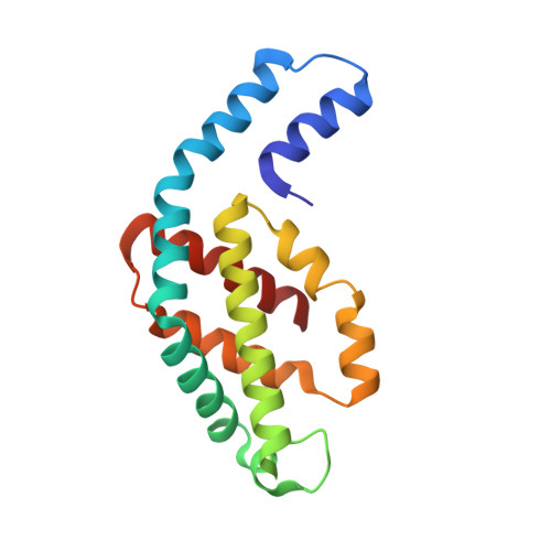
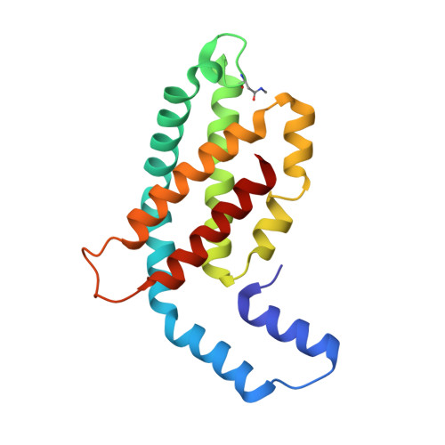
1KTP

Crystal structure of c-phycocyanin of synechococcus vulcanus at 1.6 angstroms


Experimental Data Snapshot

  • Method: X-RAY DIFFRACTION
  • Resolution: 1.60 Å
  • R-Value Free: 0.247 
  • R-Value Work: 0.216 
  • R-Value Observed: 0.216 

Starting Model: experimental
View more details

wwPDB Validation   3D Report Full Report


Ligand Structure Quality Assessment 


This is version 1.3 of the entry. See complete history


Literature

Refined structure of c-phycocyanin from the cyanobacterium Synechococcus vulcanus at 1.6 A: insights into the role of solvent molecules in thermal stability and co-factor structure

Adir, N.Vainer, R.Lerner, N.

(2002) Biochim Biophys Acta 1556: 168-174

  • DOI: https://doi.org/10.1016/s0005-2728(02)00359-6
  • Primary Citation of Related Structures:  
    1KTP

  • PubMed Abstract: 

    The crystal structure of the light-harvesting phycobiliprotein, c-phycocyanin from the thermophilic cyanobacterium Synechococcus vulcanus has been refined to 1.6 A resolution based on the previously determined lower resolution structure (PDB entry 1I7Y). The improved data was collected using synchrotron radiation at 100 K. The significantly improved crystallographic data has lead to improved calculated electron density maps, allowing the unambiguous positioning of all protein and co-factor atoms and the positioning of 377 solvent molecules. The positions of solvent molecules at specific sites important for stabilization of different levels of self-assembly of the phycobilisome structure were identified and the bonding network is described. The presence of solvent molecules in the vicinity of the co-factors and in intermolecular spaces is identified and their possible roles are suggested. All three of the phycocyanobilin co-factors bind water molecules at specific sites between the propionic acid side chains. Molecular dynamic (MD) simulations support that these special waters have a role in stabilization of this conformation. On the basis of the crystal packing reported here and in comparison to other phycobiliprotein crystal forms, we have analyzed the roles of specific sites on the formation of the phycobilisome complex.


  • Organizational Affiliation

    Department of Chemistry and Institute of Catalysis, Science and Technology, Technion-Israel Institute of Technology, City, Haifa 32000, Technion, Israel. nadir@tx.technion.ac.il


Macromolecules
Find similar proteins by:  (by identity cutoff)  |  3D Structure
Entity ID: 1
MoleculeChains Sequence LengthOrganismDetailsImage
C-PHYCOCYANIN ALPHA SUBUNIT162Thermostichus vulcanusMutation(s): 0 
UniProt
Find proteins for P50032 (Thermosynechococcus vestitus (strain NIES-2133 / IAM M-273 / BP-1))
Explore P50032 
Go to UniProtKB:  P50032
Entity Groups  
Sequence Clusters30% Identity50% Identity70% Identity90% Identity95% Identity100% Identity
UniProt GroupP50032
Sequence Annotations
Expand
  • Reference Sequence
Find similar proteins by:  (by identity cutoff)  |  3D Structure
Entity ID: 2
MoleculeChains Sequence LengthOrganismDetailsImage
C-PHYCOCYANIN BETA SUBUNIT172Thermostichus vulcanusMutation(s): 1 
UniProt
Find proteins for P50033 (Thermosynechococcus vestitus (strain NIES-2133 / IAM M-273 / BP-1))
Explore P50033 
Go to UniProtKB:  P50033
Entity Groups  
Sequence Clusters30% Identity50% Identity70% Identity90% Identity95% Identity100% Identity
UniProt GroupP50033
Sequence Annotations
Expand
  • Reference Sequence
Small Molecules
Modified Residues  1 Unique
IDChains TypeFormula2D DiagramParent
MEN
Query on MEN
B
L-PEPTIDE LINKINGC5 H10 N2 O3ASN
Experimental Data & Validation

Experimental Data

  • Method: X-RAY DIFFRACTION
  • Resolution: 1.60 Å
  • R-Value Free: 0.247 
  • R-Value Work: 0.216 
  • R-Value Observed: 0.216 
  • Space Group: H 3 2
Unit Cell:
Length ( Å )Angle ( ˚ )
a = 186.346α = 90
b = 186.346β = 90
c = 59.26γ = 120
Software Package:
Software NamePurpose
DENZOdata reduction
SCALEPACKdata scaling
AMoREphasing
CNSrefinement

Structure Validation

View Full Validation Report


Ligand Structure Quality Assessment 


Entry History 

Deposition Data

Revision History  (Full details and data files)

  • Version 1.0: 2002-03-06
    Type: Initial release
  • Version 1.1: 2008-04-27
    Changes: Version format compliance
  • Version 1.2: 2011-07-13
    Changes: Version format compliance
  • Version 1.3: 2023-08-16
    Changes: Data collection, Database references, Derived calculations, Refinement description