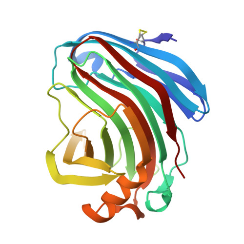
Determination of the structure of an endoglucanase from Aspergillus niger and its mode of inhibition by palladium chloride.
Khademi, S., Zhang, D., Swanson, S.M., Wartenberg, A., Witte, K., Meyer, E.F.(2002) Acta Crystallogr D Biol Crystallogr 58: 660-667
- PubMed: 11914491
- DOI: https://doi.org/10.1107/s0907444902003360
- Primary Citation of Related Structures:
1KS4, 1KS5 - PubMed Abstract:
The fungus Aspergillus niger is a main source of industrial cellulase. beta-1,4-Endoglucanase is the major component of cellulase from A. niger. In spite of widespread applications, little is known about the structure of this enzyme. Here, the structure of beta-1,4-endoglucanase from A. niger (EglA) was determined at 2.1 A resolution. Although there is a low sequence identity between EglA and CelB2, another member of family 12, the three-dimensional structures of their core regions are quite similar. The structural differences are mostly found in the loop regions, where CelB2 has an extra beta-sheet (beta-sheet C) at the non-reducing end of the binding cleft of the native enzyme. Incubation of EglA with PdCl(2) irreversibly inhibits the EglA activity. Structural studies of the enzyme-palladium complex show that three Pd(2+) ions bind to each EglA molecule. One of the Pd(2+) ions forms a coordinate covalent bond with Met118 S(delta) and the nucleophilic Glu116 O(epsilon1) at the active site of the enzyme. The other two Pd(2+) ions bind on the surface of the protein. Binding of Pd(2+) ions to EglA does not change the general conformation of the backbone of the protein significantly. Based on this structural study, one can conclude that the palladium ion directly binds to and blocks the active site of EglA and thus inactivates the enzyme.
- Biographics Laboratory, Texas A&M University, Department of Biochemistry and Biophysics, College Station, TX 77843-2128, USA.
Organizational Affiliation:


