1KOJ

Crystal structure of rabbit phosphoglucose isomerase complexed with 5-phospho-D-arabinonohydroxamic acid


Experimental Data Snapshot

  • Method: X-RAY DIFFRACTION
  • Resolution: 1.90 Å
  • R-Value Free: 0.254 
  • R-Value Work: 0.220 
  • R-Value Observed: 0.223 

wwPDB Validation   3D Report Full Report


Ligand Structure Quality Assessment 


This is version 1.3 of the entry. See complete history


Literature

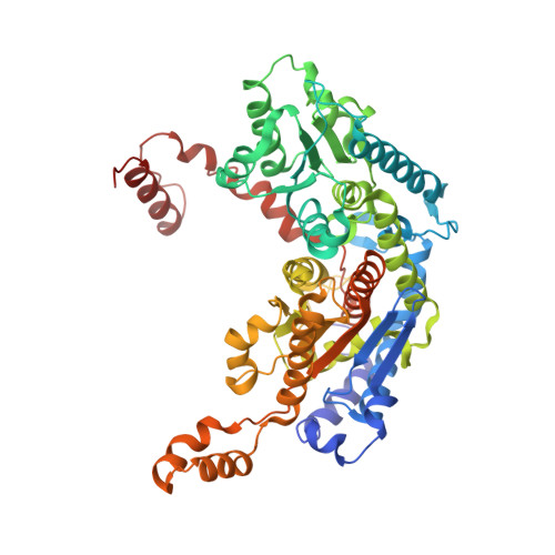
The crystal structure of rabbit phosphoglucose isomerase complexed with 5-phospho-D-arabinonohydroxamic acid.

Arsenieva, D.Hardre, R.Salmon, L.Jeffery, C.J.

(2002) Proc Natl Acad Sci U S A 99: 5872-5877

  • DOI: https://doi.org/10.1073/pnas.052131799
  • Primary Citation of Related Structures:  
    1KOJ

  • PubMed Abstract: 

    Phosphoglucose isomerase (EC ) catalyzes the second step in glycolysis, the reversible isomerization of D-glucose 6-phosphate to D-fructose 6-phosphate. The reaction mechanism involves acid-base catalysis with proton transfer and proceeds through a cis-enediol(ate) intermediate. 5-Phospho-D-arabinonohydroxamic acid (5PAH) is a synthetic small molecule that resembles the reaction intermediate, differing only in that it has a nitrogen atom in place of C1. Hence, 5PAH is the best inhibitor of the isomerization reaction reported to date with a K(i) of 2 x 10(-7) M. Here we report the crystal structure of rabbit phosphoglucose isomerase complexed with 5PAH at 1.9 A resolution. The interaction of 5PAH with amino acid residues in the enzyme active site supports a model of the catalytic mechanism in which Glu-357 transfers a proton between C1 and C2 and Arg-272 helps stabilize the intermediate. It also suggests a mechanism for proton transfer between O1 and O2.


  • Organizational Affiliation

    Laboratory for Molecular Biology, MC567, Department of Biological Sciences, University of Illinois, Chicago, IL 60607, USA.


Macromolecules
Find similar proteins by:  (by identity cutoff)  |  3D Structure
Entity ID: 1
MoleculeChains Sequence LengthOrganismDetailsImage
Glucose-6-phosphate isomerase
A, B
557Oryctolagus cuniculusMutation(s): 0 
EC: 5.3.1.9
UniProt
Find proteins for Q9N1E2 (Oryctolagus cuniculus)
Explore Q9N1E2 
Go to UniProtKB:  Q9N1E2
Entity Groups  
Sequence Clusters30% Identity50% Identity70% Identity90% Identity95% Identity100% Identity
UniProt GroupQ9N1E2
Sequence Annotations
Expand
  • Reference Sequence
Small Molecules
Ligands 1 Unique
IDChains Name / Formula / InChI Key2D Diagram3D Interactions
PAN
Query on PAN

Download Ideal Coordinates CCD File 
C [auth A],
D [auth B]
5-PHOSPHO-D-ARABINOHYDROXAMIC ACID
C5 H12 N O9 P
OHQFMJPEBPCIEQ-JJYYJPOSSA-N
Binding Affinity Annotations 
IDSourceBinding Affinity
PAN PDBBind:  1KOJ Ki: 200 (nM) from 1 assay(s)
BindingDB:  1KOJ Ki: 100 (nM) from 1 assay(s)
Binding MOAD:  1KOJ Ki: 200 (nM) from 1 assay(s)
Experimental Data & Validation

Experimental Data

  • Method: X-RAY DIFFRACTION
  • Resolution: 1.90 Å
  • R-Value Free: 0.254 
  • R-Value Work: 0.220 
  • R-Value Observed: 0.223 
  • Space Group: C 2 2 21
Unit Cell:
Length ( Å )Angle ( ˚ )
a = 82.69α = 90
b = 115.97β = 90
c = 271.85γ = 90
Software Package:
Software NamePurpose
CNSrefinement
DENZOdata reduction
SCALEPACKdata scaling
CNSphasing

Structure Validation

View Full Validation Report


Ligand Structure Quality Assessment 


Entry History 

Deposition Data

Revision History  (Full details and data files)

  • Version 1.0: 2002-05-03
    Type: Initial release
  • Version 1.1: 2008-04-27
    Changes: Version format compliance
  • Version 1.2: 2011-07-13
    Changes: Version format compliance
  • Version 1.3: 2023-08-16
    Changes: Data collection, Database references, Derived calculations, Refinement description