Structure-based enzyme inhibitor design: modeling studies and crystal structure analysis of Pneumocystis carinii dihydrofolate reductase ternary complex with PT653 and NADPH.
Cody, V., Galitsky, N., Luft, J.R., Pangborn, W., Rosowsky, A., Queener, S.F.(2002) Acta Crystallogr D Biol Crystallogr 58: 946-954
- PubMed: 12037296 Search on PubMed
- DOI: https://doi.org/10.1107/s090744490200505x
- Primary Citation Related Structures:
1KLK - PubMed Abstract:
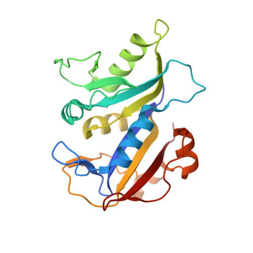
Structural data are reported for N-(2,4-diaminopteridin-6-yl)methyldibenz[b,f]azepine (PT653), an example of structure-based inhibitor design with 21-fold selectivity for Pneumocystis carinii dihydrofolate reductase (pcDHFR) relative to rat liver dihydrofolate reductase (rlDHFR). These data test the hypothesis that 2,4-diaminopteridines with a bulky N,N-diarylaminomethyl side chain at the 6-position could fit better into the larger active site of pcDHFR than into that of mammalian DHFR. The crystal structure of the ternary complex of NADPH, PT653 and pcDHFR, refined to 2.4 A resolution, reveals that PT653 binds in a different orientation than predicted from modeling studies reported previously [Rosowsky et al. (1999), J. Med. Chem. 42, 4853-4860]. These crystal data show that the pteridine-ring plane is tilted compared with that observed in the crystal structure of the pcDHFR methotrexate (MTX) NADPH ternary complex used as a template to model PT653 binding. Also, as a result of this tilt, the dibenzoazepine ring is bound deeper into the p-aminobenzoyl folate binding pocket of pcDHFR, thereby relieving close intermolecular contacts predicted from the modeling data. By far the most significant structural change, but more subtle in magnitude, is the ligand-induced conformational shift of 1.2 A away from the inhibitor of residues 61-66 in helix C. The other major effect is the unwinding of the short helical segment involving loop 47 which has a different conformation to that observed in other pcDHFR complexes [Cody et al. (1999), Biochemistry, 38, 4303-4312]. The favorable pcDHFR selectivity of PT653 could be a result of ligand-induced fit of the large hydrophobic dibenzazepine ring which occupies regions of the enzyme active site not probed by other antifolates and which take advantage of sequence and conformational differences between the structures of human and pcDHFR. These data suggest that such hydrophobic analogs could be used as lead compounds in the design of more pcDHFR-selective antifolates. Enzyme inhibition data also show that PT653 is 102-fold selective for Toxoplasma gondii (tg) DHFR relative to rlDHFR. Homology-modeling studies of the tgDHFR structure suggest that differences in ligand-binding orientation and enzyme sequence could influence the enhanced selectivity of PT653 for tgDHFR.
- Hauptman-Woodward Medical Research Institute, Inc., 73 High Street, Buffalo, NY 14203, USA. cody@hwi.buffalo.edu
Organizational Affiliation: