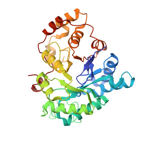
The structure of apo and holo forms of xylose reductase, a dimeric aldo-keto reductase from Candida tenuis.
Kavanagh, K.L., Klimacek, M., Nidetzky, B., Wilson, D.K.(2002) Biochemistry 41: 8785-8795
- PubMed: 12102621
- DOI: https://doi.org/10.1021/bi025786n
- Primary Citation of Related Structures:
1JEZ, 1K8C - PubMed Abstract:
Xylose reductase is a homodimeric oxidoreductase dependent on NADPH or NADH and belongs to the largely monomeric aldo-keto reductase superfamily of proteins. It catalyzes the first step in the assimilation of xylose, an aldose found to be a major constituent monosaccharide of renewable plant hemicellulosic material, into yeast metabolic pathways. It does this by reducing open chain xylose to xylitol, which is reoxidized to xylulose by xylitol dehydrogenase and metabolically integrated via the pentose phosphate pathway. No structure has yet been determined for a xylose reductase, a dimeric aldo-keto reductase or a family 2 aldo-keto reductase. The structures of the Candida tenuis xylose reductase apo- and holoenzyme, which crystallize in spacegroup C2 with different unit cells, have been determined to 2.2 A resolution and an R-factor of 17.9 and 20.8%, respectively. Residues responsible for mediating the novel dimeric interface include Asp-178, Arg-181, Lys-202, Phe-206, Trp-313, and Pro-319. Alignments with other superfamily members indicate that these interactions are conserved in other dimeric xylose reductases but not throughout the remainder of the oligomeric aldo-keto reductases, predicting alternate modes of oligomerization for other families. An arrangement of side chains in a catalytic triad shows that Tyr-52 has a conserved function as a general acid. The loop that folds over the NAD(P)H cosubstrate is disordered in the apo form but becomes ordered upon cosubstrate binding. A slow conformational isomerization of this loop probably accounts for the observed rate-limiting step involving release of cosubstrate. Xylose binding (K(m) = 87 mM) is mediated by interactions with a binding pocket that is more polar than a typical aldo-keto reductase. Modeling of xylose into the active site of the holoenzyme using ordered waters as a guide for sugar hydroxyls suggests a convincing mode of substrate binding.
- Section of Molecular and Cellular Biology, University of California-Davis, One Shields Avenue, Davis, CA 95616, USA.
Organizational Affiliation:


