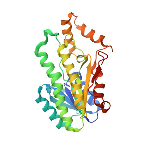
1K2W

Crystal structure of sorbitol dehydrogenase from R. sphaeroides


Experimental Data Snapshot

  • Method: X-RAY DIFFRACTION
  • Resolution: 2.40 Å
  • R-Value Free: 0.238 
  • R-Value Work: 0.188 

Starting Model: experimental
View more details

wwPDB Validation   3D Report Full Report


This is version 1.5 of the entry. See complete history


Literature

Structure of zinc-independent sorbitol dehydrogenase from Rhodobacter sphaeroides at 2.4 A resolution.

Philippsen, A.Schirmer, T.Stein, M.A.Giffhorn, F.Stetefeld, J.

(2005) Acta Crystallogr D Biol Crystallogr 61: 374-379

  • DOI: https://doi.org/10.1107/S0907444904034390
  • Primary Citation of Related Structures:  
    1K2W

  • PubMed Abstract: 

    Recombinant sorbitol dehydrogenase (SDH) from Rhodobacter sphaeroides has been crystallized in the absence of the cofactor NAD(H) and its structure determined to 2.4 A resolution using molecular replacement (refined R and R free factors of 18.8 and 23.8%, respectively). As expected from the sequence and shown by the conserved fold, SDH can be assigned to the short-chain dehydrogenase/reductase protein family. The cofactor NAD and the substrate sorbitol have been modelled into the structure and the active-site architecture, which displays the highly conserved catalytic tetrad of Asn-Ser-Tyr-Lys residues, is discussed in relation to the enzyme mechanism. This is the first structure of a bacterial SDH belonging to the SDR family.


  • Organizational Affiliation

    Division of Structural Biology, Biozentrum, University of Basel, Klingelbergstrasse 70, 4056 Basel, Switzerland. ansgar.philippsen@unibas.ch


Macromolecules
Find similar proteins by:  (by identity cutoff)  |  3D Structure
Entity ID: 1
MoleculeChains Sequence LengthOrganismDetailsImage
SORBITOL DEHYDROGENASE
A, B
256Cereibacter sphaeroidesMutation(s): 0 
EC: 1.1.1.14 (PDB Primary Data), 1.1.1 (UniProt), 1.1.1.16 (UniProt)
UniProt
Find proteins for Q59787 (Cereibacter sphaeroides)
Explore Q59787 
Go to UniProtKB:  Q59787
Entity Groups  
Sequence Clusters30% Identity50% Identity70% Identity90% Identity95% Identity100% Identity
UniProt GroupQ59787
Sequence Annotations
Expand
  • Reference Sequence
Experimental Data & Validation

Experimental Data

  • Method: X-RAY DIFFRACTION
  • Resolution: 2.40 Å
  • R-Value Free: 0.238 
  • R-Value Work: 0.188 
  • Space Group: P 31 2 1
Unit Cell:
Length ( Å )Angle ( ˚ )
a = 67.465α = 90
b = 67.465β = 90
c = 191.044γ = 120
Software Package:
Software NamePurpose
MOSFLMdata reduction
TRUNCATEdata reduction
AMoREphasing
CNSrefinement
CCP4data scaling

Structure Validation

View Full Validation Report


Entry History 

Deposition Data

Revision History  (Full details and data files)

  • Version 1.0: 2003-11-11
    Type: Initial release
  • Version 1.1: 2008-04-27
    Changes: Version format compliance
  • Version 1.2: 2011-07-13
    Changes: Version format compliance
  • Version 1.3: 2017-10-25
    Changes: Advisory, Author supporting evidence
  • Version 1.4: 2018-04-04
    Changes: Data collection
  • Version 1.5: 2023-08-16
    Changes: Advisory, Data collection, Database references, Refinement description