Siah ubiquitin ligase is structurally related to TRAF and modulates TNF-alpha signaling.
Polekhina, G., House, C.M., Traficante, N., Mackay, J.P., Relaix, F., Sassoon, D.A., Parker, M.W., Bowtell, D.D.(2002) Nat Struct Biol 9: 68-75
- PubMed: 11742346 Search on PubMed
- DOI: https://doi.org/10.1038/nsb743
- Primary Citation Related Structures:
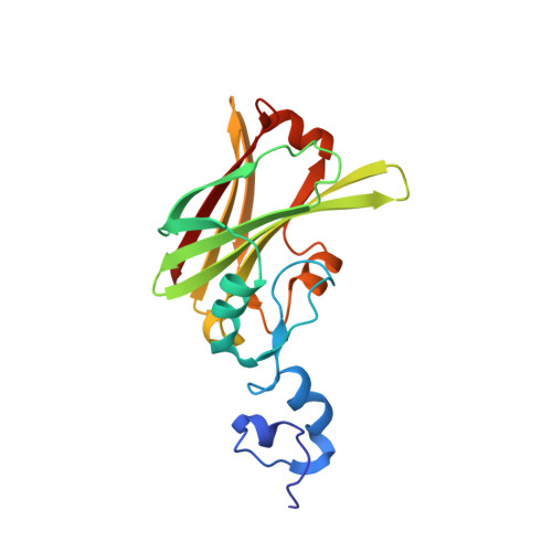
1K2F - PubMed Abstract:
Members of the Siah (seven in absentia homolog) family of RING domain proteins are components of E3 ubiquitin ligase complexes that catalyze ubiquitination of proteins. We have determined the crystal structure of the substrate-binding domain (SBD) of murine Siah1a to 2.6 A resolution. The structure reveals that Siah is a dimeric protein and that the SBD adopts an eight-stranded beta-sandwich fold that is highly similar to the TRAF-C region of TRAF (TNF-receptor associated factor) proteins. The TRAF-C region interacts with TNF-alpha receptors and TNF-receptor associated death-domain (TRADD) proteins; however, our findings indicate that these interactions are unlikely to be mimicked by Siah. The Siah structure also reveals two novel zinc fingers in a region with sequence similarity to TRAF. We find that the Siah1a SBD potentiates TNF-alpha-mediated NF-kappa B activation. Therefore, Siah proteins share important similarities with the TRAF family of proteins, including their overall domain architecture, three-dimensional structure and functional activity.
- Biota Structural Biology Laboratory, St. Vincent's Institute of Medical Research, 41 Victoria Parade, Fitzroy, Victoria 3065, Australia.
Organizational Affiliation: