


1K0Z | pdb_00001k0z
1K0Z | pdb_00001k0z
Crystal Structure of the PvuII endonuclease with Pr3+ and SO4 ions bound in the active site at 2.05A.
- PDB DOI: https://doi.org/10.2210/pdb1K0Z/pdb
- Classification: HYDROLASE
- Organism(s): Proteus vulgaris
- Expression System: Escherichia coli
- Mutation(s): No
- Deposited: 2001-09-21 Released: 2003-06-17
Experimental Data Snapshot
- Method: X-RAY DIFFRACTION
- Resolution: 2.05 Å
- R-Value Free: 0.236 (Depositor)
- R-Value Work: 0.190 (Depositor)
Starting Model: experimental
View more details
wwPDB Validation 3D Report Full Report

This is version 1.4 of the entry. See complete history.

Macromolecules
Find similar proteins by:
| 3D Structure
Entity ID: 1 | |||||
|---|---|---|---|---|---|
| Molecule | Chains | Sequence Length | Organism | Details | Image |
| Type II restriction enzyme PvuII | 156 | Proteus vulgaris | Mutation(s): 0 Gene Names: PvuiiR EC: 3.1.21.4 | 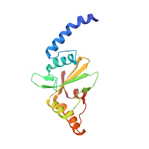 | |
UniProt | |||||
Entity Groups | |||||
| Sequence Clusters | 30% Identity50% Identity70% Identity90% Identity95% Identity100% Identity | ||||
| UniProt Group | P23657 | ||||
Sequence AnnotationsExpand | |||||
Reference Sequence | |||||
Small Molecules
| Ligands 2 Unique | |||||
|---|---|---|---|---|---|
| ID | Chains | Name / Formula / InChI Key | 2D Diagram | 3D Interactions | |
| PR Download:Ideal Coordinates CCD File | C [auth A], E [auth B] | PRASEODYMIUM ION Pr WCWKKSOQLQEJTE-UHFFFAOYSA-N |  | ||
| SO4 Download:Ideal Coordinates CCD File | D [auth A], F [auth B] | SULFATE ION O4 S QAOWNCQODCNURD-UHFFFAOYSA-L |  | ||
Experimental Data & Validation
Experimental Data
- Method: X-RAY DIFFRACTION
- Resolution: 2.05 Å
- R-Value Free: 0.236 (Depositor)
- R-Value Work: 0.190 (Depositor)
Space Group: P 21 21 2
Unit Cell:
| Length ( Å ) | Angle ( ˚ ) |
|---|---|
| a = 84.74 | α = 90 |
| b = 105.63 | β = 90 |
| c = 46.9 | γ = 90 |
| Software Name | Purpose |
|---|---|
| CNS | refinement |
| SCALA | data scaling |
| XTALVIEW | refinement |
| MOSFLM | data reduction |
| CCP4 | data scaling |
Entry History
Deposition Data
- Released Date: 2003-06-17 Deposition Author(s): Spyridaki, A., Athanasiadis, A., Matzen, C., Lanio, T., Jeltsch, A., Simoncsits, A., Scheuring-Vanamee, E., Kokkinidis, M., Pingoud, A.
Revision History (Full details and data files)
- Version 1.0: 2003-06-17
Type: Initial release - Version 1.1: 2008-04-27
Changes: Version format compliance - Version 1.2: 2011-07-13
Changes: Version format compliance - Version 1.3: 2019-07-24
Changes: Data collection, Refinement description - Version 1.4: 2023-08-16
Changes: Data collection, Database references, Derived calculations, Refinement description


