1K0Y

X-ray Crystallographic Analyses of Symmetrical Allosteric Effectors of Hemoglobin. Compounds Designed to Link Primary and Secondary Binding Sites


Experimental Data Snapshot

  • Method: X-RAY DIFFRACTION
  • Resolution: 1.87 Å
  • R-Value Free: 0.189 
  • R-Value Work: 0.163 
  • R-Value Observed: 0.164 

wwPDB Validation   3D Report Full Report


Ligand Structure Quality Assessment 


This is version 1.3 of the entry. See complete history


Literature

X-ray crystallographic analyses of symmetrical allosteric effectors of hemoglobin: compounds designed to link primary and secondary binding sites.

Safo, M.K.Boyiri, T.Burnett, J.C.Danso-Danquah, R.Moure, C.M.Joshi, G.S.Abraham, D.J.

(2002) Acta Crystallogr D Biol Crystallogr 58: 634-644

  • DOI: https://doi.org/10.1107/s0907444902002627
  • Primary Citation of Related Structures:  
    1K0Y

  • PubMed Abstract: 

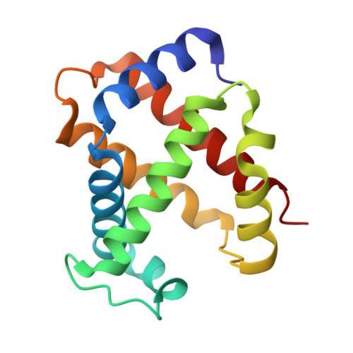
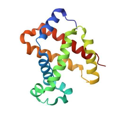
    The rational design and X-ray crystallographic analyses of two symmetrical allosteric effectors of hemoglobin (Hb) are reported. Compound design was directed by the previously solved co-crystal structure of one of the most potent allosteric effectors of Hb, 2-[4-[(3,5-dichlorophenylcarbamoyl)-methyl]-phenoxy]-2-methylpropionic acid (RSR4), which revealed two distinct binding sites for this compound in the Hb central water cavity. The primary binding site has been observed for all compounds of this structural class, which stabilize deoxy Hb by engaging in inter-dimer contacts with three of the four protein subunits. Interactions at the secondary binding site of RSR4 occur primarily between the beta(1) and beta(2) subunits and serve to further constrain the deoxy state. Based on these observations, it was hypothesized that compounds with the ability to simultaneously span and link both of these sites would possess increased potency, but at a lower molar concentration than RSR4. Two symmetrical compounds were designed and synthesized based on this hypothesis. The symmetrical effector approach was taken to minimize the number of compound orientations needed to successfully bind at either of the distinct allosteric sites. X-ray crystallographic analyses of these two effectors in complex with Hb revealed that they successfully spanned the RSR4 primary and secondary binding sites. However, the designed compounds interacted with the secondary binding site in such a way that intra-dimer, as opposed to inter-dimer, interactions were generated. In agreement with these observations, in vitro evaluation of the symmetrical effectors in Hb solution indicated that neither compound possessed the potency of RSR4. A detailed analysis of symmetrical effector-Hb contacts and comparisons with the binding contacts of RSR4 are discussed.


  • Organizational Affiliation

    Department of Medicinal Chemistry, School of Pharmacy and Institute for Structural Biology and Drug Discovery, Virginia Commonwealth University, Richmond, Virginia 23298-0540, USA. msafo@hsc.vcu.edu


Macromolecules
Find similar proteins by:  (by identity cutoff)  |  3D Structure
Entity ID: 1
MoleculeChains Sequence LengthOrganismDetailsImage
hemoglobin alpha chain
A, C
141Homo sapiensMutation(s): 0 
UniProt & NIH Common Fund Data Resources
Find proteins for P69905 (Homo sapiens)
Explore P69905 
Go to UniProtKB:  P69905
PHAROS:  P69905
Entity Groups  
Sequence Clusters30% Identity50% Identity70% Identity90% Identity95% Identity100% Identity
UniProt GroupP69905
Sequence Annotations
Expand
  • Reference Sequence
Find similar proteins by:  (by identity cutoff)  |  3D Structure
Entity ID: 2
MoleculeChains Sequence LengthOrganismDetailsImage
hemoglobin beta chain
B, D
146Homo sapiensMutation(s): 0 
UniProt & NIH Common Fund Data Resources
Find proteins for P68871 (Homo sapiens)
Explore P68871 
Go to UniProtKB:  P68871
PHAROS:  P68871
GTEx:  ENSG00000244734 
Entity Groups  
Sequence Clusters30% Identity50% Identity70% Identity90% Identity95% Identity100% Identity
UniProt GroupP68871
Sequence Annotations
Expand
  • Reference Sequence
Small Molecules
Ligands 3 Unique
IDChains Name / Formula / InChI Key2D Diagram3D Interactions
HEM
Query on HEM

Download Ideal Coordinates CCD File 
E [auth A],
G [auth B],
H [auth C],
J [auth D]
PROTOPORPHYRIN IX CONTAINING FE
C34 H32 Fe N4 O4
KABFMIBPWCXCRK-RGGAHWMASA-L
CNO
Query on CNO

Download Ideal Coordinates CCD File 
K [auth D],
L [auth D]
2-{4-[(3{2-[4-(1-CARBOXY-1-METHYL-ETHOXY)-PHENYL]-ACETYLAMINO}-PHENYLCARBAMOYL)-METHYL]-PHENOXY}-2-METHYL-PROPIONIC ACID
C30 H32 N2 O8
QRJRPWUABHMWAG-UHFFFAOYSA-N
SO4
Query on SO4

Download Ideal Coordinates CCD File 
F [auth B],
I [auth D]
SULFATE ION
O4 S
QAOWNCQODCNURD-UHFFFAOYSA-L
Experimental Data & Validation

Experimental Data

  • Method: X-RAY DIFFRACTION
  • Resolution: 1.87 Å
  • R-Value Free: 0.189 
  • R-Value Work: 0.163 
  • R-Value Observed: 0.164 
  • Space Group: P 1 21 1
Unit Cell:
Length ( Å )Angle ( ˚ )
a = 63.28α = 90
b = 83.79β = 99.33
c = 53.8γ = 90
Software Package:
Software NamePurpose
X-PLORmodel building
X-PLORrefinement
bioteXdata reduction
CCP4data scaling
X-PLORphasing

Structure Validation

View Full Validation Report


Ligand Structure Quality Assessment 


Entry History 

Deposition Data

Revision History  (Full details and data files)

  • Version 1.0: 2001-10-03
    Type: Initial release
  • Version 1.1: 2008-04-27
    Changes: Version format compliance
  • Version 1.2: 2011-07-13
    Changes: Version format compliance
  • Version 1.3: 2023-08-16
    Changes: Data collection, Database references, Derived calculations, Refinement description