1K08 | pdb_00001k08

Crystallographic Binding Study of 10 mM N-benzoyl-N'-beta-D-glucopyranosyl urea to glycogen phosphorylase b


Experimental Data Snapshot

  • Method: X-RAY DIFFRACTION
  • Resolution: 2.26 Å
  • R-Value Free: 
    0.212 (Depositor) 
  • R-Value Work: 
    0.180 (Depositor) 

Starting Model: experimental
View more details

wwPDB Validation 3D Report Full Report

Validation slider image for 1K08

This is version 1.4 of the entry. See complete history

Literature

Binding of N-acetyl-N '-beta-D-glucopyranosyl urea and N-benzoyl-N '-beta-D-glucopyranosyl urea to glycogen phosphorylase b: kinetic and crystallographic studies.

Oikonomakos, N.G.Kosmopoulou, M.Zographos, S.E.Leonidas, D.D.Chrysina, E.D.Somsak, L.Nagy, V.Praly, J.P.Docsa, T.Toth, B.Gergely, P.

(2002) Eur J Biochem 269: 1684-1696

  • DOI: https://doi.org/10.1046/j.1432-1327.2002.02813.x
  • Primary Citation Related Structures: 
    1K06, 1K08, 1KTI

  • PubMed Abstract: 

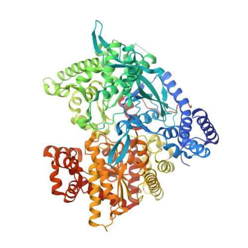
    Two substituted ureas of beta-D-glucose, N-acetyl-N'-beta-D-glucopyranosyl urea (Acurea) and N-benzoyl-N'-beta-D-glucopyranosyl urea (Bzurea), have been identified as inhibitors of glycogen phosphorylase, a potential target for therapeutic intervention in type 2 diabetes. To elucidate the structural basis of inhibition, we determined the structure of muscle glycogen phosphorylase b (GPb) complexed with the two compounds at 2.0 A and 1.8 A resolution, respectively. The structure of the GPb-Acurea complex reveals that the inhibitor can be accommodated in the catalytic site of T-state GPb with very little change in the tertiary structure. The glucopyranose moiety makes the standard hydrogen bonds and van der Waals contacts as observed in the GPb-glucose complex, while the acetyl urea moiety is in a favourable electrostatic environment and makes additional polar contacts with the protein. The structure of the GPb-Bzurea complex shows that Bzurea binds tightly at the catalytic site and induces substantial conformational changes in the vicinity of the catalytic site. In particular, the loop of the polypeptide chain containing residues 282-287 shifts 1.3-3.7 A (Calpha atoms) to accommodate Bzurea. Bzurea can also occupy the new allosteric site, some 33 A from the catalytic site, which is currently the target for the design of antidiabetic drugs.


  • Organizational Affiliation
    • Institute of Biological Research and Biotechnology, The National Hellenic Research Foundation, Athens, Greece. ngo@eie.gr

Macromolecule Content 

  • Total Structure Weight: 98.19 kDa 
  • Atom Count: 6,953 
  • Modeled Residue Count: 817 
  • Deposited Residue Count: 842 
  • Unique protein chains: 1

Macromolecules

Find similar proteins by:|  3D Structure
Entity ID: 1
MoleculeChains  Sequence LengthOrganismDetailsImage
Glycogen Phosphorylase842Oryctolagus cuniculusMutation(s): 0 
EC: 2.4.1.1
UniProt
Find proteins for P00489 (Oryctolagus cuniculus)
Explore P00489 
Go to UniProtKB:  P00489
Entity Groups
Sequence Clusters30% Identity50% Identity70% Identity90% Identity95% Identity100% Identity
UniProt GroupP00489
Sequence Annotations
Expand
Reference Sequence

Small Molecules

Ligands 2 Unique
IDChains Name / Formula / InChI Key2D Diagram3D Interactions
BZD

Query on BZD


Download:Ideal Coordinates CCD File
B [auth A],
C [auth A]
N-[(phenylcarbonyl)carbamoyl]-beta-D-glucopyranosylamine
C14 H18 N2 O7
JSBCZGSPFATCOV-BZNQNGANSA-N
PLP

Query on PLP


Download:Ideal Coordinates CCD File
D [auth A]PYRIDOXAL-5'-PHOSPHATE
C8 H10 N O6 P
NGVDGCNFYWLIFO-UHFFFAOYSA-N

Experimental Data & Validation

Experimental Data

  • Method: X-RAY DIFFRACTION
  • Resolution: 2.26 Å
  • R-Value Free:  0.212 (Depositor) 
  • R-Value Work:  0.180 (Depositor) 
Space Group: P 43 21 2
Unit Cell:
Length ( Å )Angle ( ˚ )
a = 128.519α = 90
b = 128.519β = 90
c = 116.497γ = 90
Software Package:
Software NamePurpose
X-PLORmodel building
X-PLORrefinement
DENZOdata reduction
SCALEPACKdata scaling
X-PLORphasing

Structure Validation

View Full Validation Report


Entry History 

Revision History  (Full details and data files)

  • Version 1.0: 2001-10-03
    Type: Initial release
  • Version 1.1: 2008-04-27
    Changes: Version format compliance
  • Version 1.2: 2011-07-13
    Changes: Derived calculations, Version format compliance
  • Version 1.3: 2020-07-29
    Type: Remediation
    Reason: Carbohydrate remediation
    Changes: Data collection, Derived calculations, Structure summary
  • Version 1.4: 2023-08-16
    Changes: Data collection, Database references, Refinement description, Structure summary