Cell Transformation by the v-myc Oncogene Abrogates c-Myc/Max-mediated Suppression of a C/EBPbeta-dependent Lipocalin Gene.
Hartl, M., Matt, T., Schueler, W., Siemeister, G., Kontaxis, G., Kloiber, K., Konrat, R., Bister, K.(2003) J Mol Biology 333: 33-46
- PubMed: 14516741 Search on PubMed
- DOI: https://doi.org/10.1016/j.jmb.2003.08.018
- Primary Citation Related Structures:
1JZU - PubMed Abstract:
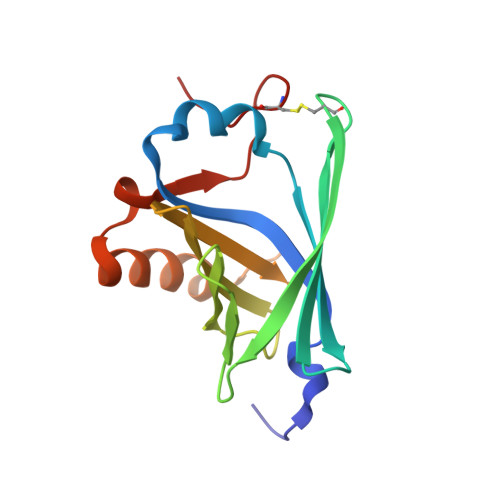
Using differential hybridization techniques, a cDNA clone (Q83) was isolated that corresponds to a highly abundant mRNA in quail embryo fibroblasts transformed by the v-myc oncogene. The deduced 178 amino acid protein product of Q83 contains an N-terminal signal sequence and a lipocalin sequence motif, the hallmark of a family of secretory proteins binding and transporting small hydrophobic molecules of diverse biological function, including retinoids and steroids. The quail Q83 protein displays 87% sequence identity with a developmentally regulated chicken protein, termed p20K or Ch21. Cell transformation specifically by v-myc, but not by other oncogenic agents, induces high-level expression of Q83 mRNA and of the Q83 protein. Nucleotide sequence analysis and transcriptional mapping revealed that the Q83 gene encompasses seven exons with the coding region confined to exons 1 through 6. The promoter region contains consensus binding sites for the transcriptional regulators Myc and C/EBP beta. Transcriptional activation of Q83 is principally dependent on C/EBP beta, but is blocked in normal cells by the endogenous c-Myc/Max/Mad transcription factor network. In v-myc-transformed cells, high-level expression of the v-Myc protein and formation of highly stable v-Myc/Max heterodimers leads to abrogation of Q83 gene suppression and activation by C/EBP beta. A 157 amino acid residue recombinant protein representing the secreted form of Q83 was used for structure determination by nuclear magnetic resonance spectroscopy. Q83 folds into a single globular domain of the lipocalin-type. The central part consists of an eight-stranded up-and-down beta-barrel core flanked by an N-terminal 3(10)-like helix and a C-terminal alpha-helix. The orientation of the C-terminal alpha-helix is partially determined by a disulfide bridge between Cys59 and Cys152. The three-dimensional structure determination of the Q83 protein will facilitate the identification of its authentic ligand and the assessment of its biological function, including the putative role in myc-induced cell transformation.
- Institute of Biochemistry, University of Innsbruck, A-6020 Innsbruck, Austria.
Organizational Affiliation: