1JYA | pdb_00001jya

Crystal Structure of SycE


Experimental Data Snapshot

  • Method: X-RAY DIFFRACTION
  • Resolution: 1.74 Å
  • R-Value Free: 
    0.266 (Depositor), 0.271 (DCC) 
  • R-Value Work: 
    0.236 (Depositor), 0.242 (DCC) 

wwPDB Validation 3D Report Full Report

Validation slider image for 1JYA

This is version 1.4 of the entry. See complete history

Literature

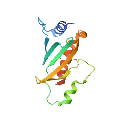
Structure of the Yersinia type III secretory system chaperone SycE.

Birtalan, S.Ghosh, P.

(2001) Nat Struct Biol 8: 974-978

  • DOI: https://doi.org/10.1038/nsb1101-974
  • Primary Citation Related Structures: 
    1JYA

  • PubMed Abstract: 

    In the type III secretory system of bacterial pathogens, a large number of sequence-divergent but characteristically small (approximately 14-19 kDa), acidic (pI approximately 4-5) chaperone proteins have been identified. We present the 1.74 A resolution crystal structure of the Yersinia pseudotuberculosis chaperone SycE, whose action in promoting translocation of YopE into host macrophages is essential to Yersinia pathogenesis. SycE, a compact, globular dimer with a novel fold, has two large hydrophobic surface patches that may form binding sites for YopE or other type III components. These patches are formed by structurally key residues that are conserved among many chaperones, suggesting shared structural and functional relationships. A negative electrostatic potential covers almost the entire surface of SycE and is likely conserved in character, but not in detail, among chaperones. The structure provides the first structural insights into possible modes of action of SycE and type III chaperones in general.


  • Organizational Affiliation
    • Department of Chemistry and Biochemistry, University of California at San Diego, La Jolla, California 92093-0314, USA.

Macromolecule Content 

  • Total Structure Weight: 29.6 kDa 
  • Atom Count: 2,028 
  • Modeled Residue Count: 238 
  • Deposited Residue Count: 260 
  • Unique protein chains: 1

Macromolecules

Find similar proteins by:|  3D Structure
Entity ID: 1
MoleculeChains  Sequence LengthOrganismDetailsImage
YOPE regulator
A, B
130Yersinia pseudotuberculosisMutation(s): 0 
Gene Names: syce
UniProt
Find proteins for P31491 (Yersinia pestis)
Explore P31491 
Go to UniProtKB:  P31491
Entity Groups
Sequence Clusters30% Identity50% Identity70% Identity90% Identity95% Identity100% Identity
UniProt GroupP31491
Sequence Annotations
Expand
Reference Sequence

Small Molecules

Modified Residues  1 Unique
IDChains TypeFormula2D DiagramParent
MSE
Query on MSE
A, B
L-PEPTIDE LINKINGC5 H11 N O2 SeMET

Experimental Data & Validation

Experimental Data

  • Method: X-RAY DIFFRACTION
  • Resolution: 1.74 Å
  • R-Value Free:  0.266 (Depositor), 0.271 (DCC) 
  • R-Value Work:  0.236 (Depositor), 0.242 (DCC) 
Space Group: C 2 2 21
Unit Cell:
Length ( Å )Angle ( ˚ )
a = 46α = 90
b = 75.19β = 90
c = 137.74γ = 90
Software Package:
Software NamePurpose
DENZOdata reduction
SCALEPACKdata scaling
SOLVEphasing
SHARPphasing
CNSrefinement

Structure Validation

View Full Validation Report


Entry History 

Deposition Data

Revision History  (Full details and data files)

  • Version 1.0: 2001-10-31
    Type: Initial release
  • Version 1.1: 2008-04-27
    Changes: Version format compliance
  • Version 1.2: 2011-07-13
    Changes: Version format compliance
  • Version 1.3: 2018-01-31
    Changes: Advisory, Experimental preparation
  • Version 1.4: 2024-10-30
    Changes: Advisory, Data collection, Database references, Derived calculations, Structure summary