


Examination of the Structural Basis for O(H) Blood Group Specificity by Ulex europaeus Lectin I
Audette, G.F., Olson, D.J.H., Ross, A.R.S., Quail, J.W., Delbaere, L.T.J.(2002) Can J Chem 80: 1010-1021
Experimental Data Snapshot
Starting Model: experimental
View more details
(2002) Can J Chem 80: 1010-1021
Entity ID: 1 | |||||
|---|---|---|---|---|---|
| Molecule | Chains | Sequence Length | Organism | Details | Image |
| anti-H(O) lectin I | 242 | Ulex europaeus | Mutation(s): 0 | 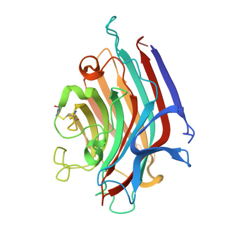 | |
UniProt | |||||
Entity Groups | |||||
| Sequence Clusters | 30% Identity50% Identity70% Identity90% Identity95% Identity100% Identity | ||||
| UniProt Group | P22972 | ||||
Sequence AnnotationsExpand | |||||
Reference Sequence | |||||
| Ligands 4 Unique | |||||
|---|---|---|---|---|---|
| ID | Chains | Name / Formula / InChI Key | 2D Diagram | 3D Interactions | |
| MFU Download:Ideal Coordinates CCD File | E [auth A], H [auth B], K [auth C], O [auth D] | methyl alpha-L-fucopyranoside C7 H14 O5 OHWCAVRRXKJCRB-CXNFULCWSA-N |  | ||
| MRD Download:Ideal Coordinates CCD File | N [auth C] | (4R)-2-METHYLPENTANE-2,4-DIOL C6 H14 O2 SVTBMSDMJJWYQN-RXMQYKEDSA-N |  | ||
| MN Download:Ideal Coordinates CCD File | F [auth A], I [auth B], L [auth C], P [auth D] | MANGANESE (II) ION Mn WAEMQWOKJMHJLA-UHFFFAOYSA-N |  | ||
| CA Download:Ideal Coordinates CCD File | G [auth A], J [auth B], M [auth C], Q [auth D] | CALCIUM ION Ca BHPQYMZQTOCNFJ-UHFFFAOYSA-N |  | ||
| Length ( Å ) | Angle ( ˚ ) |
|---|---|
| a = 71.81 | α = 90 |
| b = 69 | β = 106.76 |
| c = 119.02 | γ = 90 |
| Software Name | Purpose |
|---|---|
| DENZO | data reduction |
| SCALEPACK | data scaling |
| AMoRE | phasing |
| X-PLOR | refinement |


