1JWP | pdb_00001jwp

Structure of M182T mutant of TEM-1 beta-lactamase


Experimental Data Snapshot

  • Method: X-RAY DIFFRACTION
  • Resolution: 1.75 Å
  • R-Value Free: 
    0.220 (Depositor), 0.222 (DCC) 
  • R-Value Work: 
    0.186 (Depositor), 0.190 (DCC) 

Starting Model: experimental
View more details

wwPDB Validation 3D Report Full Report

Validation slider image for 1JWP

This is version 1.5 of the entry. See complete history

Literature

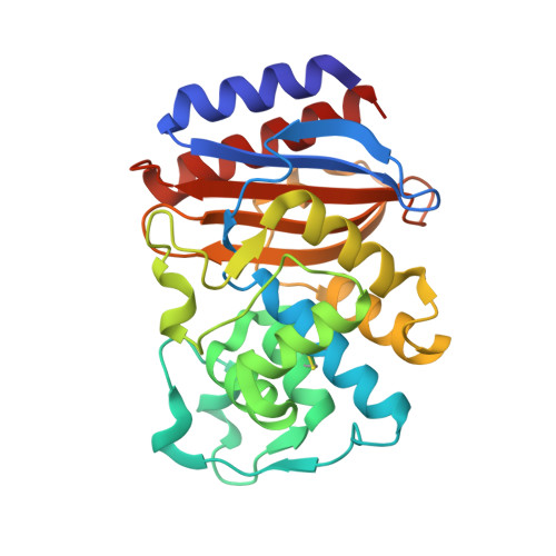
Evolution of an antibiotic resistance enzyme constrained by stability and activity trade-offs.

Wang, X.Minasov, G.Shoichet, B.K.

(2002) J Mol Biology 320: 85-95

  • DOI: https://doi.org/10.1016/S0022-2836(02)00400-X
  • Primary Citation Related Structures: 
    1JWP, 1JWV, 1JWZ

  • PubMed Abstract: 

    Pressured by antibiotic use, resistance enzymes have been evolving new activities. Does such evolution have a cost? To investigate this question at the molecular level, clinically isolated mutants of the beta-lactamase TEM-1 were studied. When purified, mutant enzymes had increased activity against cephalosporin antibiotics but lost both thermodynamic stability and kinetic activity against their ancestral targets, penicillins. The X-ray crystallographic structures of three mutant enzymes were determined. These structures suggest that activity gain and stability loss is related to an enlarged active site cavity in the mutant enzymes. In several clinically isolated mutant enzymes, a secondary substitution is observed far from the active site (Met182-->Thr). This substitution had little effect on enzyme activity but restored stability lost by substitutions near the active site. This regained stability conferred an advantage in vivo. This pattern of stability loss and restoration may be common in the evolution of new enzyme activity.


  • Organizational Affiliation
    • Department of Molecular Pharmacology and Biological Chemistry, Northwestern University School of Medicine, 303 East Chicago Avenue, Chicago, IL 60611-3008, USA.

Macromolecule Content 

  • Total Structure Weight: 29.01 kDa 
  • Atom Count: 2,322 
  • Modeled Residue Count: 263 
  • Deposited Residue Count: 263 
  • Unique protein chains: 1

Macromolecules

Find similar proteins by:|  3D Structure
Entity ID: 1
MoleculeChains  Sequence LengthOrganismDetailsImage
BETA-LACTAMASE TEM263Escherichia coliMutation(s): 1 
Gene Names: bla
EC: 3.5.2.6
UniProt
Find proteins for P62593 (Escherichia coli)
Explore P62593 
Go to UniProtKB:  P62593
Entity Groups
Sequence Clusters30% Identity50% Identity70% Identity90% Identity95% Identity100% Identity
UniProt GroupP62593
Sequence Annotations
Expand
Reference Sequence

Small Molecules

Ligands 1 Unique
IDChains Name / Formula / InChI Key2D Diagram3D Interactions
PO4

Query on PO4


Download:Ideal Coordinates CCD File
B [auth A]PHOSPHATE ION
O4 P
NBIIXXVUZAFLBC-UHFFFAOYSA-K

Experimental Data & Validation

Experimental Data

  • Method: X-RAY DIFFRACTION
  • Resolution: 1.75 Å
  • R-Value Free:  0.220 (Depositor), 0.222 (DCC) 
  • R-Value Work:  0.186 (Depositor), 0.190 (DCC) 
Space Group: P 21 21 21
Unit Cell:
Length ( Å )Angle ( ˚ )
a = 41.465α = 90
b = 61.691β = 90
c = 89.205γ = 90
Software Package:
Software NamePurpose
DENZOdata reduction
SCALEPACKdata scaling
AMoREphasing
CNSrefinement

Structure Validation

View Full Validation Report


Entry History 

Deposition Data

Revision History  (Full details and data files)

  • Version 1.0: 2002-06-05
    Type: Initial release
  • Version 1.1: 2008-04-27
    Changes: Version format compliance
  • Version 1.2: 2011-07-13
    Changes: Version format compliance
  • Version 1.3: 2021-10-27
    Changes: Database references, Derived calculations
  • Version 1.4: 2023-08-16
    Changes: Data collection, Refinement description
  • Version 1.5: 2024-10-30
    Changes: Structure summary