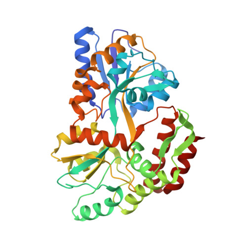
1JVX

Maltodextrin-binding protein variant D207C/A301GS/P316C cross-linked in crystal


Experimental Data Snapshot

  • Method: X-RAY DIFFRACTION
  • Resolution: 2.50 Å
  • R-Value Free: 0.304 
  • R-Value Work: 0.232 
  • R-Value Observed: 0.239 

Starting Model: experimental
View more details

wwPDB Validation   3D Report Full Report


This is version 2.2 of the entry. See complete history


Literature

Crystine: Fibrous Biomolecular Material from Protein Crystals Cross-linked in a Specific Geometry

Srinivasan, U.Iyer, G.H.Przybycien, T.A.Samsonoff, W.A.Bell, J.A.

(2002) Protein Eng 15: 895-902

  • DOI: https://doi.org/10.1093/protein/15.11.895
  • Primary Citation of Related Structures:  
    1JVX, 1JVY

  • PubMed Abstract: 

    Cysteine substitutions were engineered on the surface of maltose binding protein to produce crystine fibers, linear polymers of folded protein formed within a crystal. Disulfide bond formation between adjacent protein molecules within the lattice was monitored by X-ray crystallography. The cross-linked crystals were resistant to dissolution in water or neutral buffer solutions, even though the cross-linking was one-dimensional. However, crystine fibers were observed by transmission electron microscopy to dissociate from the crystals in acidic solutions. Some fibers remained associated as two-dimensional bundles or sheets, with a repeat unit along the fibers consistent with the packing of the individual protein molecules in the crystal. Neutralization of the acidic solutions caused the fibers to re-associate as a solid. Crystine threads were drawn out of this solution. In scanning electron microscopy images, many individual fibers could be seen unwinding from the ends of some threads. Crystine fibers are a new type of biomolecular material with potential applications wherever the use of proteins in a fibrous form is desirable, for example, the incorporation of enzymes into cloth or filtration material.


  • Organizational Affiliation

    Department of Chemistry, Rensselaer Polytechnic Institute, Troy, NY 12180, USA.


Macromolecules
Find similar proteins by:  (by identity cutoff)  |  3D Structure
Entity ID: 1
MoleculeChains Sequence LengthOrganismDetailsImage
maltodextrin-binding protein372Escherichia coliMutation(s): 2 
Gene Names: malE
UniProt
Find proteins for P0AEX9 (Escherichia coli (strain K12))
Explore P0AEX9 
Go to UniProtKB:  P0AEX9
Entity Groups  
Sequence Clusters30% Identity50% Identity70% Identity90% Identity95% Identity100% Identity
UniProt GroupP0AEX9
Sequence Annotations
Expand
  • Reference Sequence
Oligosaccharides

Help

Entity ID: 2
MoleculeChains Length2D Diagram Glycosylation3D Interactions
alpha-D-glucopyranose-(1-4)-alpha-D-glucopyranose
B
2N/A
Glycosylation Resources
GlyTouCan:  G07411ON
GlyCosmos:  G07411ON
Biologically Interesting Molecules (External Reference) 1 Unique
Experimental Data & Validation

Experimental Data

  • Method: X-RAY DIFFRACTION
  • Resolution: 2.50 Å
  • R-Value Free: 0.304 
  • R-Value Work: 0.232 
  • R-Value Observed: 0.239 
  • Space Group: C 1 2 1
Unit Cell:
Length ( Å )Angle ( ˚ )
a = 103.61α = 90
b = 67.58β = 113.31
c = 57.27γ = 90
Software Package:
Software NamePurpose
MOSFLMdata reduction
SCALAdata scaling
CNSrefinement
CNXrefinement
CCP4data scaling
CNSphasing

Structure Validation

View Full Validation Report


Entry History 

Deposition Data

Revision History  (Full details and data files)

  • Version 1.0: 2001-09-12
    Type: Initial release
  • Version 1.1: 2008-04-27
    Changes: Version format compliance
  • Version 1.2: 2011-07-13
    Changes: Version format compliance
  • Version 2.0: 2020-07-29
    Type: Remediation
    Reason: Carbohydrate remediation
    Changes: Atomic model, Data collection, Derived calculations, Non-polymer description, Structure summary
  • Version 2.1: 2021-10-27
    Changes: Database references, Structure summary
  • Version 2.2: 2023-08-16
    Changes: Data collection, Refinement description