1JSY

Crystal structure of bovine arrestin-2


Experimental Data Snapshot

  • Method: X-RAY DIFFRACTION
  • Resolution: 2.90 Å
  • R-Value Free: 0.238 
  • R-Value Work: 0.230 
  • R-Value Observed: 0.227 

wwPDB Validation   3D Report Full Report


This is version 1.3 of the entry. See complete history


Literature

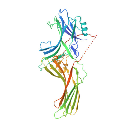
Scaffolding functions of arrestin-2 revealed by crystal structure and mutagenesis.

Milano, S.K.Pace, H.C.Kim, Y.M.Brenner, C.Benovic, J.L.

(2002) Biochemistry 41: 3321-3328

  • DOI: https://doi.org/10.1021/bi015905j
  • Primary Citation of Related Structures:  
    1JSY

  • PubMed Abstract: 

    Arrestin binding to activated, phosphorylated G protein-coupled receptors (GPCRs) represents a critical step in regulation of light- and hormone-dependent signaling. Nonvisual arrestins, such as arrestin-2, interact with multiple proteins for the purpose of propagating and terminating signaling events. Using a combination of X-ray crystallography, molecular modeling, mutagenesis, and binding analysis, we reveal structural features of arrestin-2 that may enable simultaneous binding to phosphorylated receptor, SH3 domains, phosphoinositides, and beta-adaptin. The structure of full-length arrestin-2 thus provides a uniquely oriented scaffold for assembly of multiple, diverse molecules involved in GPCR signal transduction.


  • Organizational Affiliation

    Structural Biology and Bioinformatics Program, Kimmel Cancer Center, Thomas Jefferson University, Philadelphia, PA 19107, USA.


Macromolecules
Find similar proteins by:  (by identity cutoff)  |  3D Structure
Entity ID: 1
MoleculeChains Sequence LengthOrganismDetailsImage
Bovine arrestin-2 (full length)418Bos taurusMutation(s): 0 
UniProt
Find proteins for P17870 (Bos taurus)
Explore P17870 
Go to UniProtKB:  P17870
Entity Groups  
Sequence Clusters30% Identity50% Identity70% Identity90% Identity95% Identity100% Identity
UniProt GroupP17870
Sequence Annotations
Expand
  • Reference Sequence
Experimental Data & Validation

Experimental Data

  • Method: X-RAY DIFFRACTION
  • Resolution: 2.90 Å
  • R-Value Free: 0.238 
  • R-Value Work: 0.230 
  • R-Value Observed: 0.227 
  • Space Group: P 32 2 1
Unit Cell:
Length ( Å )Angle ( ˚ )
a = 78.952α = 90
b = 78.952β = 90
c = 158.924γ = 120
Software Package:
Software NamePurpose
AMoREphasing
CNSrefinement
DENZOdata reduction
SCALEPACKdata scaling

Structure Validation

View Full Validation Report


Entry History 

Deposition Data

Revision History  (Full details and data files)

  • Version 1.0: 2002-03-27
    Type: Initial release
  • Version 1.1: 2008-04-27
    Changes: Version format compliance
  • Version 1.2: 2011-07-13
    Changes: Version format compliance
  • Version 1.3: 2023-08-16
    Changes: Data collection, Database references, Refinement description