1JR9

Crystal Structure of manganese superoxide dismutases from Bacillus halodenitrificans


Experimental Data Snapshot

  • Method: X-RAY DIFFRACTION
  • Resolution: 2.80 Å
  • R-Value Free: 0.289 
  • R-Value Work: 0.220 

wwPDB Validation   3D Report Full Report


This is version 1.2 of the entry. See complete history


Literature

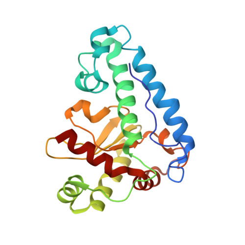
Three-dimensional structure of manganese superoxide dismutase from Bacillus halodenitrificans, a component of the so-called "green protein".

Liao, J.Liu, M.Y.Chang, T.Li, M.Le Gall, J.Gui, L.L.Zhang, J.P.Jiang, T.Liang, D.C.Chang, W.R.

(2002) J Struct Biol 139: 171-180

  • DOI: https://doi.org/10.1016/s1047-8477(02)00531-2
  • Primary Citation of Related Structures:  
    1JR9

  • PubMed Abstract: 
  • A so-called "green protein" has been purified from a moderate halophilic eubacterium, Bacillus halodenitrificans (ATCC 49067), under anaerobic conditions. The protein, which might play an important role in denitrification, dissociates mainly into two components after exposure to air: a manganese superoxide dismutase (GP-MnSOD) and a nucleoside diphosphate kinase ...

    A so-called "green protein" has been purified from a moderate halophilic eubacterium, Bacillus halodenitrificans (ATCC 49067), under anaerobic conditions. The protein, which might play an important role in denitrification, dissociates mainly into two components after exposure to air: a manganese superoxide dismutase (GP-MnSOD) and a nucleoside diphosphate kinase. As a first step in elucidating the overall structure of the green protein and the role of each component, the 2.8-A resolution crystal structure of GP-MnSOD was determined. Compared with other manganese dismutases, GP-MnSOD shows two significant characteristics. The first is that the entrance to its substrate channel has an additional basic residue-Lys38. The second is that its surface is decorated with an excess of acidic over basic residues. All these structural features may be related to GP-MnSOD's high catalytic activity and its endurance against the special cytoplasm of B. halodenitrificans. The structure of GP-MnSOD provides the basis for recognizing its possible role and assembly state in the green protein.


    Organizational Affiliation

    National Laboratory of Biomacromolecules, Institute of Biophysics, Chinese Academy of Sciences, 15 Datun Road, Chaoyang District, Beijing, China.


Macromolecules
Find similar proteins by:  (by identity cutoff)  |  3D Structure
Entity ID: 1
MoleculeChainsSequence LengthOrganismDetailsImage
manganese superoxide dismutase202Virgibacillus halodenitrificansMutation(s): 0 
Gene Names: sodA
EC: 1.15.1.1
UniProt
Find proteins for Q7SIC3 (Virgibacillus halodenitrificans)
Explore Q7SIC3 
Go to UniProtKB:  Q7SIC3
Entity Groups  
Sequence Clusters30% Identity50% Identity70% Identity90% Identity95% Identity100% Identity
UniProt GroupQ7SIC3
Protein Feature View
Expand
  • Reference Sequence
Small Molecules
Ligands 2 Unique
IDChainsName / Formula / InChI Key2D Diagram3D Interactions
ZN
Query on ZN

Download Ideal Coordinates CCD File 
C [auth A]ZINC ION
Zn
PTFCDOFLOPIGGS-UHFFFAOYSA-N
 Ligand Interaction
MN
Query on MN

Download Ideal Coordinates CCD File 
B [auth A]MANGANESE (II) ION
Mn
WAEMQWOKJMHJLA-UHFFFAOYSA-N
 Ligand Interaction
Experimental Data & Validation

Experimental Data

  • Method: X-RAY DIFFRACTION
  • Resolution: 2.80 Å
  • R-Value Free: 0.289 
  • R-Value Work: 0.220 
  • Space Group: P 43 21 2
Unit Cell:
Length ( Å )Angle ( ˚ )
a = 93.12α = 90
b = 93.12β = 90
c = 63.527γ = 90
Software Package:
Software NamePurpose
DENZOdata reduction
SCALEPACKdata scaling
AMoREphasing
CNSrefinement

Structure Validation

View Full Validation Report


Entry History 

Deposition Data

Revision History  (Full details and data files)

  • Version 1.0: 2002-08-14
    Type: Initial release
  • Version 1.1: 2007-10-24
    Changes: Version format compliance
  • Version 1.2: 2011-07-13
    Changes: Version format compliance