Life on carbon monoxide: X-ray structure of Rhodospirillum rubrum Ni-Fe-S carbon monoxide dehydrogenase.
Drennan, C.L., Heo, J., Sintchak, M.D., Schreiter, E., Ludden, P.W.(2001) Proc Natl Acad Sci U S A 98: 11973-11978
- PubMed: 11593006 Search on PubMedSearch on PubMed Central
- DOI: https://doi.org/10.1073/pnas.211429998
- Primary Citation Related Structures:
1JQK - PubMed Abstract:
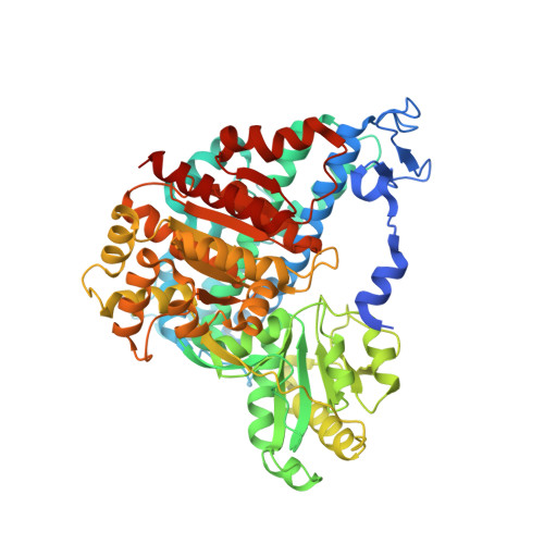
A crystal structure of the anaerobic Ni-Fe-S carbon monoxide dehydrogenase (CODH) from Rhodospirillum rubrum has been determined to 2.8-A resolution. The CODH family, for which the R. rubrum enzyme is the prototype, catalyzes the biological oxidation of CO at an unusual Ni-Fe-S cluster called the C-cluster. The Ni-Fe-S C-cluster contains a mononuclear site and a four-metal cubane. Surprisingly, anomalous dispersion data suggest that the mononuclear site contains Fe and not Ni, and the four-metal cubane has the form [NiFe(3)S(4)] and not [Fe(4)S(4)]. The mononuclear site and the four-metal cluster are bridged by means of Cys(531) and one of the sulfides of the cube. CODH is organized as a dimer with a previously unidentified [Fe(4)S(4)] cluster bridging the two subunits. Each monomer is comprised of three domains: a helical domain at the N terminus, an alpha/beta (Rossmann-like) domain in the middle, and an alpha/beta (Rossmann-like) domain at the C terminus. The helical domain contributes ligands to the bridging [Fe(4)S(4)] cluster and another [Fe(4)S(4)] cluster, the B-cluster, which is involved in electron transfer. The two Rossmann domains contribute ligands to the active site C-cluster. This x-ray structure provides insight into the mechanism of biological CO oxidation and has broader significance for the roles of Ni and Fe in biological systems.
- Department of Chemistry, Massachusetts Institute of Technology, Cambridge, MA 02139, USA. cdrennan@mit.edu
Organizational Affiliation: