1JKW | pdb_00001jkw

STRUCTURE OF CYCLIN MCS2


Experimental Data Snapshot

  • Method: X-RAY DIFFRACTION
  • Resolution: 2.60 Å
  • R-Value Free: 
    0.272 (Depositor) 
  • R-Value Work: 
    0.222 (Depositor) 
  • R-Value Observed: 
    0.222 (Depositor) 

wwPDB Validation 3D Report Full Report

Validation slider image for 1JKW

This is version 1.4 of the entry. See complete history

Literature

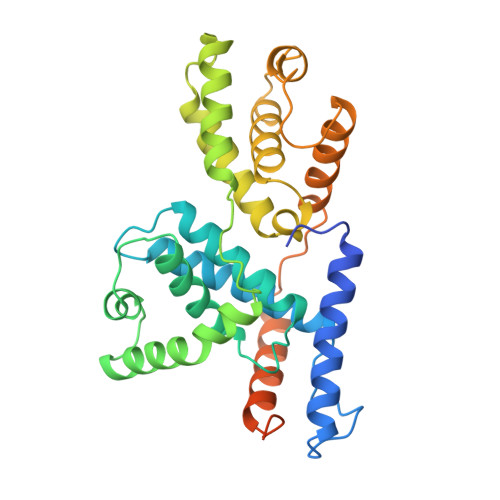
The crystal structure of human cyclin H.

Andersen, G.Poterszman, A.Egly, J.M.Moras, D.Thierry, J.C.

(1996) FEBS Lett 397: 65-69

  • DOI: https://doi.org/10.1016/s0014-5793(96)01143-x
  • Primary Citation Related Structures: 
    1JKW

  • PubMed Abstract: 

    The crystal structure of human cyclin H has been solved at 2.6 A resolution by the MIR method and refined to an R-factor of 23.1%. The core of the molecule consists of two helical repeats adopting the canonical cyclin fold already observed in the structures of cyclin A [Brown et al. (1995) Structure 3, 1235-1247; Jeffrey et al. (1995) Nature 376, 313-320; Russo et al. (1996) Nature 382, 325-331] and TFIIB [Nikoilov et al. (1995) Nature 377, 119-128]. The N-terminal and C-terminal residues form a new domain built on two long helices interacting essentially with the first repeat of the molecule.


  • Organizational Affiliation
    • Institut de Génétique et Biologie Moléculaire et Cellulaire, CNRS/INSERM/ULP, Illkirch, France.

Macromolecule Content 

  • Total Structure Weight: 37.68 kDa 
  • Atom Count: 2,299 
  • Modeled Residue Count: 277 
  • Deposited Residue Count: 323 
  • Unique protein chains: 1

Macromolecules

Find similar proteins by:|  3D Structure
Entity ID: 1
MoleculeChains  Sequence LengthOrganismDetailsImage
CYCLIN H323Homo sapiensMutation(s): 0 
Gene Names: HUMAN CYCLIN H
UniProt & NIH Common Fund Data Resources
Find proteins for P51946 (Homo sapiens)
Explore P51946 
Go to UniProtKB:  P51946
PHAROS:  P51946
GTEx:  ENSG00000134480 
Entity Groups
Sequence Clusters30% Identity50% Identity70% Identity90% Identity95% Identity100% Identity
UniProt GroupP51946
Sequence Annotations
Expand
Reference Sequence

Experimental Data & Validation

Experimental Data

  • Method: X-RAY DIFFRACTION
  • Resolution: 2.60 Å
  • R-Value Free:  0.272 (Depositor) 
  • R-Value Work:  0.222 (Depositor) 
  • R-Value Observed: 0.222 (Depositor) 
Space Group: I 41 2 2
Unit Cell:
Length ( Å )Angle ( ˚ )
a = 84.06α = 90
b = 84.06β = 90
c = 373.3γ = 90
Software Package:
Software NamePurpose
X-PLORmodel building
X-PLORrefinement
XDSdata reduction
X-PLORphasing

Structure Validation

View Full Validation Report


Entry History 

Deposition Data

Revision History  (Full details and data files)

  • Version 1.0: 1997-01-11
    Type: Initial release
  • Version 1.1: 2008-03-24
    Changes: Version format compliance
  • Version 1.2: 2011-07-13
    Changes: Version format compliance
  • Version 1.3: 2024-02-07
    Changes: Data collection, Database references, Other
  • Version 1.4: 2026-01-28
    Changes: Database references, Structure summary