1JJV

DEPHOSPHO-COA KINASE IN COMPLEX WITH ATP


Experimental Data Snapshot

  • Method: X-RAY DIFFRACTION
  • Resolution: 2.00 Å
  • R-Value Free: 0.218 
  • R-Value Work: 0.165 
  • R-Value Observed: 0.168 

wwPDB Validation   3D Report Full Report


Ligand Structure Quality Assessment 


This is version 1.3 of the entry. See complete history


Literature

Crystal structure of dephospho-coenzyme A kinase from Haemophilus influenzae.

Obmolova, G.Teplyakov, A.Bonander, N.Eisenstein, E.Howard, A.J.Gilliland, G.L.

(2001) J Struct Biol 136: 119-125

  • DOI: https://doi.org/10.1006/jsbi.2001.4428
  • Primary Citation of Related Structures:  
    1JJV

  • PubMed Abstract: 

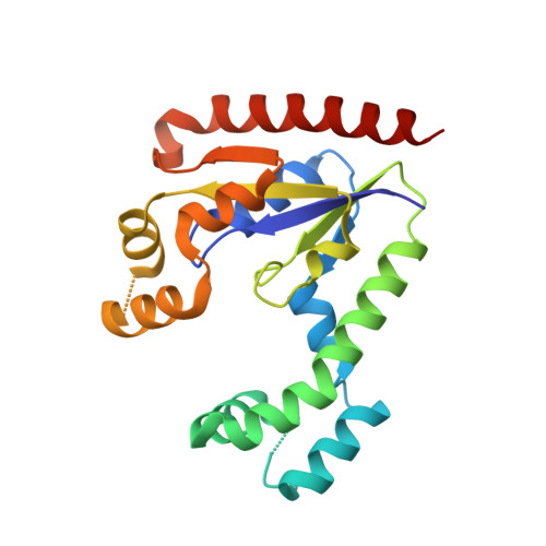
    Dephospho-coenzyme A kinase catalyzes the final step in CoA biosynthesis, the phosphorylation of the 3'-hydroxyl group of ribose using ATP as a phosphate donor. The protein from Haemophilus influenzae was cloned and expressed, and its crystal structure was determined at 2.0-A resolution in complex with ATP. The protein molecule consists of three domains: the canonical nucleotide-binding domain with a five-stranded parallel beta-sheet, the substrate-binding alpha-helical domain, and the lid domain formed by a pair of alpha-helices. The overall topology of the protein resembles the structures of nucleotide kinases. ATP binds in the P-loop in a manner observed in other kinases. The CoA-binding site is located at the interface of all three domains. The double-pocket structure of the substrate-binding site is unusual for nucleotide kinases. Amino acid residues implicated in substrate binding and catalysis have been identified. The structure analysis suggests large domain movements during the catalytic cycle.


  • Organizational Affiliation

    Center for Advanced Research in Biotechnology of the University of Maryland Biotechnology Institute, National Institute of Standards and Technology, 9600 Gudelsky Drive, Rockville, Maryland 20850, USA.


Macromolecules
Find similar proteins by:  (by identity cutoff)  |  3D Structure
Entity ID: 1
MoleculeChains Sequence LengthOrganismDetailsImage
DEPHOSPHO-COA KINASE206Haemophilus influenzaeMutation(s): 0 
Gene Names: YacE
EC: 2.7.1.24
UniProt
Find proteins for P44920 (Haemophilus influenzae (strain ATCC 51907 / DSM 11121 / KW20 / Rd))
Explore P44920 
Go to UniProtKB:  P44920
Entity Groups  
Sequence Clusters30% Identity50% Identity70% Identity90% Identity95% Identity100% Identity
UniProt GroupP44920
Sequence Annotations
Expand
  • Reference Sequence
Experimental Data & Validation

Experimental Data

  • Method: X-RAY DIFFRACTION
  • Resolution: 2.00 Å
  • R-Value Free: 0.218 
  • R-Value Work: 0.165 
  • R-Value Observed: 0.168 
  • Space Group: C 1 2 1
Unit Cell:
Length ( Å )Angle ( ˚ )
a = 126α = 90
b = 45.9β = 97.6
c = 41.7γ = 90
Software Package:
Software NamePurpose
DENZOdata reduction
SCALEPACKdata scaling
MLPHAREphasing
REFMACrefinement

Structure Validation

View Full Validation Report


Ligand Structure Quality Assessment 


Entry History 

Deposition Data

Revision History  (Full details and data files)

  • Version 1.0: 2002-05-01
    Type: Initial release
  • Version 1.1: 2008-04-27
    Changes: Version format compliance
  • Version 1.2: 2011-07-13
    Changes: Version format compliance
  • Version 1.3: 2024-02-07
    Changes: Data collection, Database references, Derived calculations