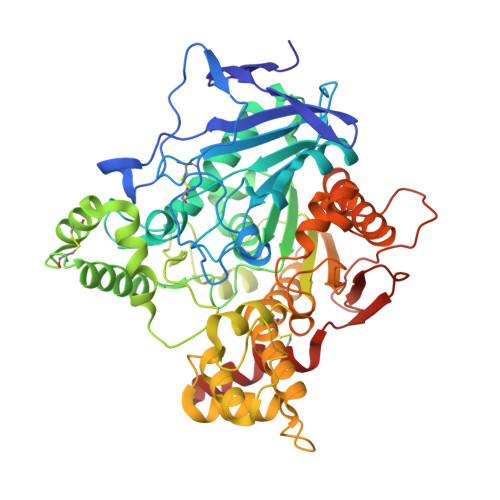
1JJB

A neutral molecule in cation-binding site: Specific binding of PEG-SH to Acetylcholinesterase from Torpedo californica


Experimental Data Snapshot

  • Method: X-RAY DIFFRACTION
  • Resolution: 2.30 Å
  • R-Value Free: 0.224 
  • R-Value Work: 0.178 
  • R-Value Observed: 0.178 

wwPDB Validation   3D Report Full Report


This is version 1.4 of the entry. See complete history


Literature

A neutral molecule in a cation-binding site: specific binding of a PEG-SH to acetylcholinesterase from Torpedo californica.

Koellner, G.Steiner, T.Millard, C.B.Silman, I.Sussman, J.L.

(2002) J Mol Biol 320: 721-725

  • DOI: https://doi.org/10.1016/s0022-2836(02)00475-8
  • Primary Citation of Related Structures:  
    1JJB

  • PubMed Abstract: 

    The crystal structure of acetylcholinesterase from Torpedo californica complexed with the uncharged inhibitor, PEG-SH-350 (containing mainly heptameric polyethylene glycol with a terminal thiol group) is determined at 2.3 A resolution. This is an untypical acetylcholinesterase inhibitor, since it lacks the cationic moiety typical of the substrate (acetylcholine). In the crystal structure, the elongated ligand extends along the whole of the deep and narrow active-site gorge, with the terminal thiol group bound near the bottom, close to the catalytic site. Unexpectedly, the cation-binding site (formed by the faces of aromatic side-chains) is occupied by CH(2) groups of the inhibitor, which are engaged in C-H...pi interactions that structurally mimic the cation-pi interactions made by the choline moiety of acetylcholine. In addition, the PEG-SH molecule makes numerous other weak but specific interactions of the C-H...O and C-H...pi types.


  • Organizational Affiliation

    Institut für Chemie-Kristallographie, Freie Universität Berlin, Takustrasse 6, D-14195 Berlin, Germany. koellner@chemie.fu-berlin.de


Macromolecules
Find similar proteins by:  (by identity cutoff)  |  3D Structure
Entity ID: 1
MoleculeChains Sequence LengthOrganismDetailsImage
ACETYLCHOLINESTERASE532Tetronarce californicaMutation(s): 0 
EC: 3.1.1.7
UniProt
Find proteins for P04058 (Tetronarce californica)
Explore P04058 
Go to UniProtKB:  P04058
Entity Groups  
Sequence Clusters30% Identity50% Identity70% Identity90% Identity95% Identity100% Identity
UniProt GroupP04058
Glycosylation
Glycosylation Sites: 2
Sequence Annotations
Expand
  • Reference Sequence
Small Molecules
Ligands 2 Unique
IDChains Name / Formula / InChI Key2D Diagram3D Interactions
PE7
Query on PE7

Download Ideal Coordinates CCD File 
D [auth A]1-DEOXY-1-THIO-HEPTAETHYLENE GLYCOL
C14 H30 O7 S
ACMBXVJDKVNCGH-UHFFFAOYSA-N
NAG
Query on NAG

Download Ideal Coordinates CCD File 
B [auth A],
C [auth A]
2-acetamido-2-deoxy-beta-D-glucopyranose
C8 H15 N O6
OVRNDRQMDRJTHS-FMDGEEDCSA-N
Experimental Data & Validation

Experimental Data

  • Method: X-RAY DIFFRACTION
  • Resolution: 2.30 Å
  • R-Value Free: 0.224 
  • R-Value Work: 0.178 
  • R-Value Observed: 0.178 
  • Space Group: P 31 2 1
Unit Cell:
Length ( Å )Angle ( ˚ )
a = 111.2α = 90
b = 111.2β = 90
c = 137.119γ = 120
Software Package:
Software NamePurpose
DENZOdata reduction
SCALEPACKdata scaling
CNSrefinement
CNSphasing

Structure Validation

View Full Validation Report


Entry History 

Deposition Data

Revision History  (Full details and data files)

  • Version 1.0: 2002-07-17
    Type: Initial release
  • Version 1.1: 2008-04-27
    Changes: Version format compliance
  • Version 1.2: 2011-07-13
    Changes: Non-polymer description, Version format compliance
  • Version 1.3: 2020-07-29
    Type: Remediation
    Reason: Carbohydrate remediation
    Changes: Data collection, Derived calculations, Structure summary
  • Version 1.4: 2021-06-02
    Changes: Derived calculations, Structure summary