1JIE | pdb_00001jie

Crystal structure of bleomycin-binding protein from bleomycin-producing Streptomyces verticillus complexed with metal-free bleomycin


Experimental Data Snapshot

  • Method: X-RAY DIFFRACTION
  • Resolution: 1.80 Å
  • R-Value Free: 
    0.218 (Depositor) 
  • R-Value Observed: 
    0.160 (Depositor) 

Starting Model: experimental
View more details

wwPDB Validation 3D Report Full Report

Validation slider image for 1JIE

This is version 1.4 of the entry. See complete history

Literature

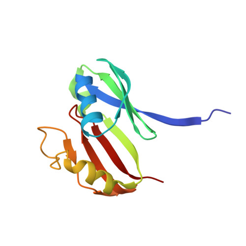
The 1.6-A crystal structure of the copper(II)-bound bleomycin complexed with the bleomycin-binding protein from bleomycin-producing Streptomyces verticillus.

Sugiyama, M.Kumagai, T.Hayashida, M.Maruyama, M.Matoba, Y.

(2002) J Biological Chem 277: 2311-2320

  • DOI: https://doi.org/10.1074/jbc.M103278200
  • Primary Citation Related Structures: 
    1JIE, 1JIF

  • PubMed Abstract: 

    Bleomycin (Bm) in the culture broth of Streptomyces verticillus is complexed with Cu(2+) (Cu(II)). In the present study, we determined the x-ray crystal structures of the Cu(II)-bound and the metal-free types of Bm at a high resolution of 1.6 and 1.8 A, respectively, which are complexed with a Bm resistance determinant from Bm-producing S. verticillus, designated BLMA. In the current model of Cu(II).Bm complexed with BLMA, two Cu(II).Bm molecules bind to the BLMA dimer. The electron density map shows that the copper ion is clearly defined in the metal-binding domain of the Bm molecule. The metal ion is penta-coordinated by a tetragonal monopyramidal cage of nitrogens and binds to the primary amine of the beta-aminoalanine moiety of Bm. The binding experiment between Bm and BLMA showed that each of the two Bm-binding pockets has a different dissociation constant (K(d)(1) and K(d)(2)). The K(d)(1) value of 630 nm for the first Bm binding is larger than the K(d)(2) value of 120 nm, indicating that the first Bm binding gives rise to a cooperative binding of the second Bm to the other pocket.


  • Organizational Affiliation
    • Institute of Pharmaceutical Sciences, Faculty of Medicine, Hiroshima University, Kasumi 1-2-3, Minami-ku, Hiroshima 734-8551, Japan. sugi@hiroshima-u.ac.jp

Macromolecule Content 

  • Total Structure Weight: 29.32 kDa 
  • Atom Count: 2,229 
  • Modeled Residue Count: 244 
  • Deposited Residue Count: 244 
  • Unique protein chains: 1

Macromolecules

Find similar proteins by:|  3D Structure
Entity ID: 1
MoleculeChains  Sequence LengthOrganismDetailsImage
bleomycin-binding protein
A, B
122Streptomyces verticillusMutation(s): 0 
Gene Names: blmA
UniProt
Find proteins for Q53793 (Streptomyces verticillus)
Explore Q53793 
Go to UniProtKB:  Q53793
Entity Groups
Sequence Clusters30% Identity50% Identity70% Identity90% Identity95% Identity100% Identity
UniProt GroupQ53793
Sequence Annotations
Expand
Reference Sequence

Small Molecules

Ligands 1 Unique
IDChains Name / Formula / InChI Key2D Diagram3D Interactions
BLM

Query on BLM


Download:Ideal Coordinates CCD File
C [auth A],
D [auth B]
BLEOMYCIN A2
C55 H85 N17 O21 S3
ZGCQRJQBETWECF-UAPAGMARSA-N

Experimental Data & Validation

Experimental Data

  • Method: X-RAY DIFFRACTION
  • Resolution: 1.80 Å
  • R-Value Free:  0.218 (Depositor) 
  • R-Value Observed: 0.160 (Depositor) 
Space Group: P 1 21 1
Unit Cell:
Length ( Å )Angle ( ˚ )
a = 35.52α = 90
b = 83.42β = 104.88
c = 43.24γ = 90
Software Package:
Software NamePurpose
X-PLORmodel building
SHELXL-97refinement
X-PLORphasing

Structure Validation

View Full Validation Report


Entry History 

Deposition Data

Revision History  (Full details and data files)

  • Version 1.0: 2002-02-06
    Type: Initial release
  • Version 1.1: 2008-04-27
    Changes: Version format compliance
  • Version 1.2: 2011-07-13
    Changes: Version format compliance
  • Version 1.3: 2017-10-04
    Changes: Refinement description
  • Version 1.4: 2023-10-25
    Changes: Data collection, Database references, Derived calculations, Refinement description