1JI5

Dlp-1 from bacillus anthracis


Experimental Data Snapshot

  • Method: X-RAY DIFFRACTION
  • Resolution: 2.50 Å
  • R-Value Free: 0.291 
  • R-Value Work: 0.226 
  • R-Value Observed: 0.226 

wwPDB Validation   3D Report Full Report


This is version 1.2 of the entry. See complete history


Literature

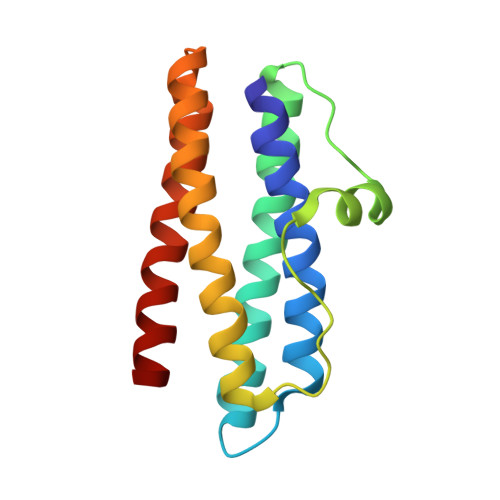
Structure of two iron-binding proteins from Bacillus anthracis.

Papinutto, E.Dundon, W.G.Pitulis, N.Battistutta, R.Montecucco, C.Zanotti, G.

(2002) J Biol Chem 277: 15093-15098

  • DOI: https://doi.org/10.1074/jbc.M112378200
  • Primary Citation of Related Structures:  
    1JI5, 1JIG

  • PubMed Abstract: 
  • Bacillus anthracis is currently under intense investigation due to its primary importance as a human pathogen. Particularly important is the development of novel anti-anthrax vaccines, devoid of the current side effects. A novel class of immunogenic bacterial proteins consists of dodecamers homologous to the DNA-binding protein of Escherichia coli (Dps) ...

    Bacillus anthracis is currently under intense investigation due to its primary importance as a human pathogen. Particularly important is the development of novel anti-anthrax vaccines, devoid of the current side effects. A novel class of immunogenic bacterial proteins consists of dodecamers homologous to the DNA-binding protein of Escherichia coli (Dps). Two Dps homologous genes are present in the B. anthracis genome. The crystal structures of these two proteins (Dlp-1 and Dlp-2) have been determined and are presented here. They are sphere-like proteins with an internal cavity. We also show that they act as ferritins and are thus involved in iron uptake and regulation, a fundamental function during bacterial growth.


    Related Citations: 
    • The Crystal Structure of Dps, a Ferritin Homolog that Binds and Protects DNA
      Grant, R.A., Filman, D.J., Finkel, S.E., Kolter, R., Hogle, J.M.
      (1998) Nat Struct Biol 5: 294
    • The Dodecameric Ferritin from Listeria innocua Contains a Novel Intersubunit Iron-binding Site
      Ilari, A., Stefanini, S., Chiancone, E., Tsernoglou, D.
      (2000) Nat Struct Biol 7: 38

    Organizational Affiliation

    Dipartimento di Chimica Organica e Centro CNR Biopolimeri, Università di Padova, Via Marzolo 1, 35131 Padova, Italy.


Macromolecules
Find similar proteins by:  (by identity cutoff)  |  3D Structure
Entity ID: 1
MoleculeChainsSequence LengthOrganismDetailsImage
Dlp-1
A, B, C, D
142Bacillus anthracisMutation(s): 0 
Gene Names: dlp-1dps2dlp1BA_5290GBAA_5290BAS4914
EC: 1.16
UniProt
Find proteins for Q8RPQ2 (Bacillus anthracis)
Explore Q8RPQ2 
Go to UniProtKB:  Q8RPQ2
Entity Groups  
Sequence Clusters30% Identity50% Identity70% Identity90% Identity95% Identity100% Identity
UniProt GroupQ8RPQ2
Protein Feature View
Expand
  • Reference Sequence
Small Molecules
Ligands 2 Unique
IDChainsName / Formula / InChI Key2D Diagram3D Interactions
MPD
Query on MPD

Download Ideal Coordinates CCD File 
G [auth B](4S)-2-METHYL-2,4-PENTANEDIOL
C6 H14 O2
SVTBMSDMJJWYQN-YFKPBYRVSA-N
 Ligand Interaction
FE
Query on FE

Download Ideal Coordinates CCD File 
E [auth A],
F [auth B],
H [auth C],
I [auth D]
FE (III) ION
Fe
VTLYFUHAOXGGBS-UHFFFAOYSA-N
 Ligand Interaction
Experimental Data & Validation

Experimental Data

  • Method: X-RAY DIFFRACTION
  • Resolution: 2.50 Å
  • R-Value Free: 0.291 
  • R-Value Work: 0.226 
  • R-Value Observed: 0.226 
  • Space Group: H 3
Unit Cell:
Length ( Å )Angle ( ˚ )
a = 89.224α = 90
b = 89.224β = 90
c = 210.165γ = 120
Software Package:
Software NamePurpose
AMoREphasing
CNSrefinement
MOSFLMdata reduction
CCP4data scaling

Structure Validation

View Full Validation Report


Entry History 

Deposition Data

Revision History  (Full details and data files)

  • Version 1.0: 2002-06-19
    Type: Initial release
  • Version 1.1: 2008-04-27
    Changes: Version format compliance
  • Version 1.2: 2011-07-13
    Changes: Derived calculations, Version format compliance