1JH4

Solution structure of the C-terminal PABC domain of human poly(A)-binding protein in complex with the peptide from Paip1


Experimental Data Snapshot

  • Method: SOLUTION NMR
  • Conformers Calculated: 200 
  • Conformers Submitted: 30 
  • Selection Criteria: structures with the lowest energy 

wwPDB Validation   3D Report Full Report


This is version 1.3 of the entry. See complete history


Literature

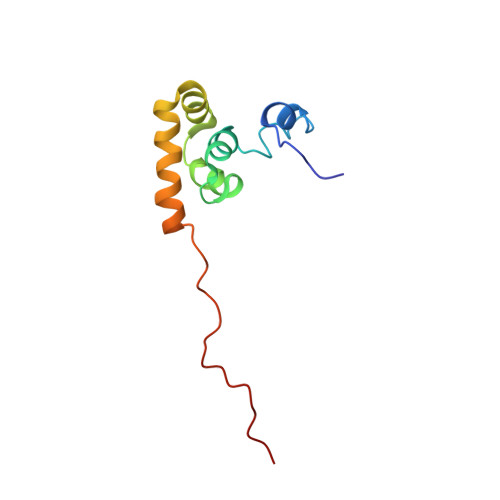
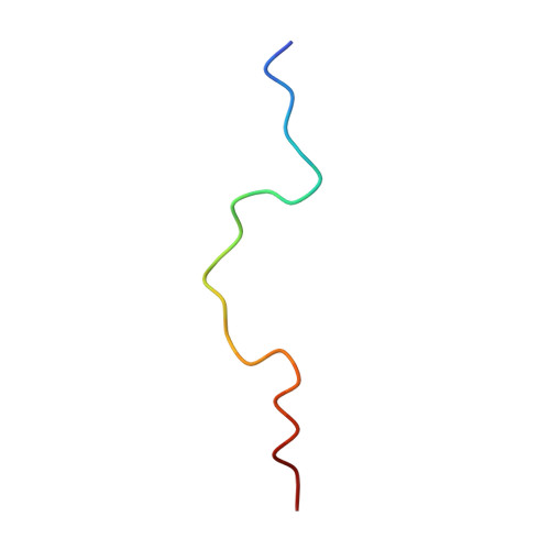
Structural basis of ligand recognition by PABC, a highly specific peptide-binding domain found in poly(A)-binding protein and a HECT ubiquitin ligase

Kozlov, G.De Crescenzo, G.Lim, N.S.Siddiqui, N.Fantus, D.Kahvejian, A.Trempe, J.F.Elias, D.Ekiel, I.Sonenberg, N.O'Connor-McCourt, M.Gehring, K.

(2004) EMBO J 23: 272-281

  • DOI: https://doi.org/10.1038/sj.emboj.7600048
  • Primary Citation of Related Structures:  
    1JGN, 1JH4

  • PubMed Abstract: 

    The C-terminal domain of poly(A)-binding protein (PABC) is a peptide-binding domain found in poly(A)-binding proteins (PABPs) and a HECT (homologous to E6-AP C-terminus) family E3 ubiquitin ligase. In protein synthesis, the PABC domain of PABP functions to recruit several translation factors possessing the PABP-interacting motif 2 (PAM2) to the mRNA poly(A) tail. We have determined the solution structure of the human PABC domain in complex with two peptides from PABP-interacting protein-1 (Paip1) and Paip2. The structures show a novel mode of peptide recognition, in which the peptide binds as a pair of beta-turns with extensive hydrophobic, electrostatic and aromatic stacking interactions. Mutagenesis of PABC and peptide residues was used to identify key protein-peptide interactions and quantified by isothermal calorimetry, surface plasmon resonance and GST pull-down assays. The results provide insight into the specificity of PABC in mediating PABP-protein interactions.


  • Organizational Affiliation

    Department of Biochemistry, McGill University, 3655 Promenade Sir William Osler, Montreal, Quebec, Canada.


Macromolecules
Find similar proteins by:  (by identity cutoff)  |  3D Structure
Entity ID: 1
MoleculeChains Sequence LengthOrganismDetailsImage
polyadenylate-binding protein 198Homo sapiensMutation(s): 0 
Gene Names: PABPC1
UniProt & NIH Common Fund Data Resources
Find proteins for P11940 (Homo sapiens)
Explore P11940 
Go to UniProtKB:  P11940
PHAROS:  P11940
GTEx:  ENSG00000070756 
Entity Groups  
Sequence Clusters30% Identity50% Identity70% Identity90% Identity95% Identity100% Identity
UniProt GroupP11940
Sequence Annotations
Expand
  • Reference Sequence

Find similar proteins by:  Sequence   |   3D Structure  

Entity ID: 2
MoleculeChains Sequence LengthOrganismDetailsImage
polyadenylate-binding protein-interacting protein-122Homo sapiensMutation(s): 0 
UniProt & NIH Common Fund Data Resources
Find proteins for Q9H074 (Homo sapiens)
Explore Q9H074 
Go to UniProtKB:  Q9H074
PHAROS:  Q9H074
GTEx:  ENSG00000172239 
Entity Groups  
Sequence Clusters30% Identity50% Identity70% Identity90% Identity95% Identity100% Identity
UniProt GroupQ9H074
Sequence Annotations
Expand
  • Reference Sequence
Experimental Data & Validation

Experimental Data

  • Method: SOLUTION NMR
  • Conformers Calculated: 200 
  • Conformers Submitted: 30 
  • Selection Criteria: structures with the lowest energy 

Structure Validation

View Full Validation Report


Entry History 

Deposition Data

Revision History  (Full details and data files)

  • Version 1.0: 2003-06-24
    Type: Initial release
  • Version 1.1: 2008-04-27
    Changes: Version format compliance
  • Version 1.2: 2011-07-13
    Changes: Version format compliance
  • Version 1.3: 2022-02-23
    Changes: Data collection, Database references, Derived calculations