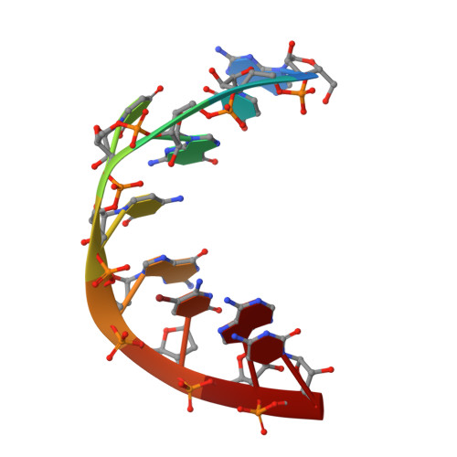
Crystal structure of an RNA duplex r(gugucgcac)(2) with uridine bulges.
Xiong, Y., Deng, J., Sudarsanakumar, C., Sundaralingam, M.(2001) J Mol Biology 313: 573-582
- PubMed: 11676540
- DOI: https://doi.org/10.1006/jmbi.2001.5045
- Primary Citation Related Structures:
1J9H - PubMed Abstract:
The crystal structure of a nonamer RNA duplex with a uridine bulge in each strand, r(gugucgcac)(2), was determined at 1.4 A resolution. The structure was solved by multiple anomalous diffraction phasing method using a three-wavelength data set collected at the Advanced Protein Source and refined to a final R(work)/R(free) of 21.2 %/23.4 % with 33,271 independent reflections (Friedel pairs unmerged). The RNA duplex crystallized in the tetragonal space group P4(1)22 with two independent molecules in the asymmetric unit. The unit cell dimensions are a=b=47.18 A and c=80.04 A. The helical region of the nonamer adopts the A-form conformation. The uridine bulges assume similar conformations, with uracils flipping out and protruding into the minor groove. The presence of the bulge induces very large twist angles (approximately +50 degrees) between the base-pairs flanking the bulges while causing profound kinks in the helix axis at the bulges. This severe twist and the large kink in turn produces a very narrow major groove at the middle of the molecule. The ribose sugars of the guanosines before the bulges adopt the C2'-endo conformation while the rest, including the bulges, are in the C3'-endo conformation. The intrastrand phosphate-phosphate (P-P) distance of the phosphate groups flanking the bulges (approximately 4.4 A) are significantly shorter than the average P-P distance in the duplex (6.0 A). This short distance between the two phosphate groups brings the non-bridging oxygen atoms close to each other where a calcium ion is bound to each strand. The calcium ions in molecule 1 are well defined while the calcium ions in molecule 2 are disordered.
- Department of Chemistry, The Ohio State University Biological Macromolecular Structure Center, 012 Rightmire Hall, 1060 Carmack Rd., Columbus, OH 43210, USA.
Organizational Affiliation: