Crystal Structure of Rat Heme Oxygenase-1 in Complex with Biliverdin-Iron Chelate: CONFORMATIONAL CHANGE OF THE DISTAL HELIX DURING THE HEME CLEAVAGE REACTION.
Sugishima, M., Sakamoto, H., Higashimoto, Y., Noguchi, M., Fukuyama, K.(2003) J Biological Chem 278: 32352-32358
- PubMed: 12794075 Search on PubMed
- DOI: https://doi.org/10.1074/jbc.M303682200
- Primary Citation Related Structures:
1J2C - PubMed Abstract:
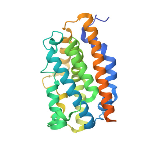
The crystal structure of rat heme oxygenase-1 in complex with biliverdin-iron chelate (biliverdin(Fe)-HO-1), the immediate precursor of the final product, biliverdin, has been determined at a 2.4-A resolution. The electron density in the heme pocket clearly showed that the tetrapyrrole ring of heme is cleaved at the alpha-meso edge. Like the heme bound to HO-1, biliverdin-iron chelate is located between the distal and proximal helices, but its accommodation state seems to be less stable in light of the disordering of the solvent-exposed propionate and vinyl groups. The middle of the distal helix is shifted away from the center of the active site in biliverdin(Fe)-HO-1, increasing the size of the heme pocket. The hydrogen-bonding interaction between Glu-29 and Gln-38, considered to restrain the orientation of the proximal helix in the heme-HO-1 complex, was lost in biliverdin(Fe)-HO-1, leading to relaxation of the helix. Biliverdin has a distorted helical conformation; the lactam oxygen atom of its pyrrole ring-A interacted with Asp-140 through a hydrogen-bonding solvent network. Because of the absence of a distal water ligand, the iron atom is five-coordinated with His-25 and four pyrrole nitrogen atoms. The coordination geometry deviates considerably from a square pyramid, suggesting that the iron may be readily dissociated. We speculate that the opened conformation of the heme pocket facilitates sequential product release, first iron then biliverdin, and that because of biliverdin's increased flexibility, iron release triggers its slow dissociation.
- Department of Biology, Graduate School of Science, Osaka University, Toyonaka, Osaka 560-0043, Japan.
Organizational Affiliation: