1J0O

High Resolution Crystal Structure of the wild type Tetraheme Cytochrome c3 from Desulfovibrio vulgaris Miyazaki F


Experimental Data Snapshot

  • Method: X-RAY DIFFRACTION
  • Resolution: 1.15 Å
  • R-Value Free: 0.167 
  • R-Value Work: 0.101 
  • R-Value Observed: 0.104 

wwPDB Validation   3D Report Full Report


This is version 1.4 of the entry. See complete history


Literature

Role of the aromatic ring of Tyr43 in tetraheme cytochrome c(3) from Desulfovibrio vulgaris Miyazaki F.

Ozawa, K.Takayama, Y.Yasukawa, F.Ohmura, T.Cusanovich, M.A.Tomimoto, Y.Ogata, H.Higuchi, Y.Akutsu, H.

(2003) Biophys J 85: 3367-3374

  • DOI: https://doi.org/10.1016/S0006-3495(03)74756-0
  • Primary Citation of Related Structures:  
    1J0O, 1J0P, 1WR5

  • PubMed Abstract: 

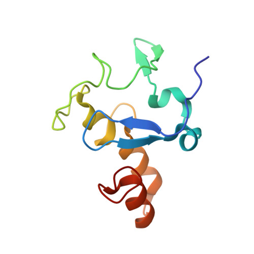
    Tyrosine 43 is positioned parallel to the fifth heme axial ligand, His34, of heme 1 in the tetraheme cytochrome c(3). The replacement of tyrosine with leucine increased the redox potential of heme 1 by 44 and 35 mV at the first and last reduction steps, respectively; its effects on the other hemes are small. In contrast, the Y43F mutation hardly changed the potentials. It shows that the aromatic ring at this position contributes to lowering the redox potential of heme 1 locally, although this cannot be the major contribution to the extremely low redox potentials of cytochrome c(3). Furthermore, temperature-dependent line-width broadening in partially reduced samples established that the aromatic ring at position 43 participates in the control of the kinetics of intramolecular electron transfer. The rate of reduction of Y43L cytochrome c(3) by 5-deazariboflavin semiquinone under partially reduced conditions was significantly different from that of the wild type in the last stage of the reduction, supporting the involvement of Tyr43 in regulation of reduction kinetics. The mutation of Y43L, however, did not induce a significant change in the crystal structure.


  • Organizational Affiliation

    Institute for Protein Research, Osaka University, Suita, Japan.


Macromolecules
Find similar proteins by:  (by identity cutoff)  |  3D Structure
Entity ID: 1
MoleculeChains Sequence LengthOrganismDetailsImage
Cytochrome c3107Nitratidesulfovibrio vulgarisMutation(s): 0 
UniProt
Find proteins for P00132 (Desulfovibrio vulgaris (strain DSM 19637 / Miyazaki F))
Explore P00132 
Go to UniProtKB:  P00132
Entity Groups  
Sequence Clusters30% Identity50% Identity70% Identity90% Identity95% Identity100% Identity
UniProt GroupP00132
Sequence Annotations
Expand
  • Reference Sequence
Small Molecules
Ligands 2 Unique
IDChains Name / Formula / InChI Key2D Diagram3D Interactions
HEM
Query on HEM

Download Ideal Coordinates CCD File 
B [auth A],
C [auth A],
D [auth A],
E [auth A]
PROTOPORPHYRIN IX CONTAINING FE
C34 H32 Fe N4 O4
KABFMIBPWCXCRK-RGGAHWMASA-L
EOH
Query on EOH

Download Ideal Coordinates CCD File 
F [auth A]
G [auth A]
H [auth A]
I [auth A]
J [auth A]
F [auth A],
G [auth A],
H [auth A],
I [auth A],
J [auth A],
K [auth A],
L [auth A],
M [auth A]
ETHANOL
C2 H6 O
LFQSCWFLJHTTHZ-UHFFFAOYSA-N
Experimental Data & Validation

Experimental Data

  • Method: X-RAY DIFFRACTION
  • Resolution: 1.15 Å
  • R-Value Free: 0.167 
  • R-Value Work: 0.101 
  • R-Value Observed: 0.104 
  • Space Group: P 21 21 21
Unit Cell:
Length ( Å )Angle ( ˚ )
a = 52.336α = 90
b = 67.347β = 90
c = 34.094γ = 90
Software Package:
Software NamePurpose
MOSFLMdata reduction
SCALAdata scaling
CNSrefinement
SHELXL-97refinement
CCP4data scaling
CNSphasing

Structure Validation

View Full Validation Report


Entry History 

Deposition Data

Revision History  (Full details and data files)

  • Version 1.0: 2003-11-19
    Type: Initial release
  • Version 1.1: 2008-04-27
    Changes: Version format compliance
  • Version 1.2: 2011-07-13
    Changes: Version format compliance
  • Version 1.3: 2019-10-23
    Changes: Data collection, Database references, Derived calculations
  • Version 1.4: 2023-12-27
    Changes: Data collection, Database references, Derived calculations