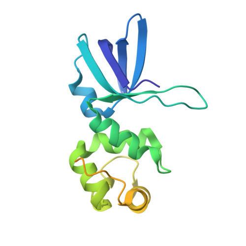
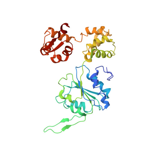
1IXR

RuvA-RuvB complex


Experimental Data Snapshot

  • Method: X-RAY DIFFRACTION
  • Resolution: 3.30 Å
  • R-Value Free: 0.335 
  • R-Value Work: 0.268 

wwPDB Validation   3D Report Full Report


Ligand Structure Quality Assessment 


This is version 1.4 of the entry. See complete history


Literature

Crystal Structure of the RuvA-RuvB Complex: A Structural Basis for the Holliday Junction Migrating Motor Machinery

Yamada, K.Miyata, T.Tsuchiya, D.Oyama, T.Fujiwara, Y.Ohnishi, T.Iwasaki, H.Shinagawa, H.Ariyoshi, M.Mayanagi, K.Morikawa, K.

(2002) Mol Cell 10: 671-681

  • DOI: https://doi.org/10.1016/s1097-2765(02)00641-x
  • Primary Citation of Related Structures:  
    1IXR, 1IXS

  • PubMed Abstract: 

    We present the X-ray structure of the RuvA-RuvB complex, which plays a crucial role in ATP-dependent branch migration. Two RuvA tetramers form the symmetric and closed octameric shell, where four RuvA domain IIIs spring out in the two opposite directions to be individually caught by a single RuvB. The binding of domain III deforms the protruding beta hairpin in the N-terminal domain of RuvB and thereby appears to induce a functional and less symmetric RuvB hexameric ring. The model of the RuvA-RuvB junction DNA ternary complex, constructed by fitting the X-ray structure into the averaged electron microscopic images of the RuvA-RuvB junction, appears to be more compatible with the branch migration mode of a fixed RuvA-RuvB interaction than with a rotational interaction mode.


  • Organizational Affiliation

    Biomolecular Engineering Research Institute, Suita, Osaka, Japan.


Macromolecules
Find similar proteins by:  (by identity cutoff)  |  3D Structure
Entity ID: 1
MoleculeChains Sequence LengthOrganismDetailsImage
Holliday junction DNA helicase ruvA
A, B
191Thermus thermophilusMutation(s): 0 
Gene Names: ruvA
UniProt
Find proteins for Q9F1Q3 (Thermus thermophilus (strain ATCC 27634 / DSM 579 / HB8))
Explore Q9F1Q3 
Go to UniProtKB:  Q9F1Q3
Entity Groups  
Sequence Clusters30% Identity50% Identity70% Identity90% Identity95% Identity100% Identity
UniProt GroupQ9F1Q3
Sequence Annotations
Expand
  • Reference Sequence
Find similar proteins by:  (by identity cutoff)  |  3D Structure
Entity ID: 2
MoleculeChains Sequence LengthOrganismDetailsImage
RuvB312Thermus thermophilusMutation(s): 1 
Gene Names: ruvB
EC: 3.6.1.3
UniProt
Find proteins for Q5SL87 (Thermus thermophilus (strain ATCC 27634 / DSM 579 / HB8))
Explore Q5SL87 
Go to UniProtKB:  Q5SL87
Entity Groups  
Sequence Clusters30% Identity50% Identity70% Identity90% Identity95% Identity100% Identity
UniProt GroupQ5SL87
Sequence Annotations
Expand
  • Reference Sequence
Small Molecules
Ligands 1 Unique
IDChains Name / Formula / InChI Key2D Diagram3D Interactions
ANP
Query on ANP

Download Ideal Coordinates CCD File 
D [auth C]PHOSPHOAMINOPHOSPHONIC ACID-ADENYLATE ESTER
C10 H17 N6 O12 P3
PVKSNHVPLWYQGJ-KQYNXXCUSA-N
Experimental Data & Validation

Experimental Data

  • Method: X-RAY DIFFRACTION
  • Resolution: 3.30 Å
  • R-Value Free: 0.335 
  • R-Value Work: 0.268 
  • Space Group: I 2 2 2
Unit Cell:
Length ( Å )Angle ( ˚ )
a = 61.581α = 90
b = 195.113β = 90
c = 246.244γ = 90
Software Package:
Software NamePurpose
DENZOdata reduction
SCALEPACKdata scaling
SOLVEphasing
CNSrefinement

Structure Validation

View Full Validation Report


Ligand Structure Quality Assessment 


Entry History 

Deposition Data

Revision History  (Full details and data files)

  • Version 1.0: 2002-11-06
    Type: Initial release
  • Version 1.1: 2008-04-27
    Changes: Version format compliance
  • Version 1.2: 2011-07-13
    Changes: Version format compliance
  • Version 1.3: 2021-11-10
    Changes: Database references, Derived calculations
  • Version 1.4: 2023-12-27
    Changes: Data collection