Enhancement of nucleoside phosphorylation activity in an acid phosphatase
Ishikawa, K., Mihara, Y., Shimba, N., Ohtsu, N., Kawasaki, H., Suzuki, E., Asano, Y.(2002) Protein Eng 15: 539-543
- PubMed: 12200535 Search on PubMed
- DOI: https://doi.org/10.1093/protein/15.7.539
- Primary Citation Related Structures:
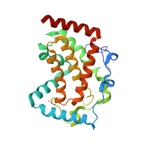
1IW8 - PubMed Abstract:
Escherichia blattae non-specific acid phosphatase (EB-NSAP) possesses a pyrophosphate-nucleoside phosphotransferase activity, which is C-5'-position selective. Current mutational and structural data were used to generate a mutant EB-NSAP for a potential industrial application as an effective and economical protein catalyst in synthesizing nucleotides from nucleosides. First, Gly74 and Ile153 were replaced by Asp and Thr, respectively, since the corresponding replacements in the homologous enzyme from Morganella morganii reduced the K(m) value for inosine and thus increased the productivity of 5'-IMP. We determined the crystal structure of G74D/I153T, which has a reduced K(m) value for inosine, as expected. The tertiary structure of G74D/I153T was virtually identical to that of the wild-type. In addition, neither of the introduced side chains of Asp74 and Thr153 is directly involved in the interaction with inosine in a hypothetical binding mode of inosine to EB-NSAP, although both residues are situated near a potential inosine-binding site. These findings suggested that a slight structural change caused by an amino acid replacement around the potential inosine-binding site could significantly reduce the K(m) value. Prompted by this hypothesis, we designed several mutations and introduced them to G74D/I153T, to decrease the K(m) value further. This strategy produced a S72F/G74D/I153T mutant with a 5.4-fold lower K(m) value and a 2.7-fold higher V(max) value as compared to the wild-type EB-NSAP.
- Central Research Laboratories, Ajinomoto Co., Inc., 1-1 Suzuki-cho Kawasaki-ku, Kawasaki 210-868, Japan.
Organizational Affiliation: