1IV6

Solution Structure of the DNA Complex of Human TRF1


Experimental Data Snapshot

  • Method: SOLUTION NMR
  • Conformers Calculated: 100 
  • Conformers Submitted: 20 
  • Selection Criteria: structures with the least restraint violations 

wwPDB Validation   3D Report Full Report


This is version 1.4 of the entry. See complete history


Literature

Solution structure of a telomeric DNA complex of human TRF1.

Nishikawa, T.Okamura, H.Nagadoi, A.Konig, P.Rhodes, D.Nishimura, Y.

(2001) Structure 9: 1237-1251

  • DOI: https://doi.org/10.1016/s0969-2126(01)00688-8
  • Primary Citation of Related Structures:  
    1ITY, 1IV6

  • PubMed Abstract: 

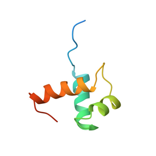
    Mammalian telomeres consist of long tandem arrays of double-stranded TTAGGG sequence motif packaged by TRF1 and TRF2. In contrast to the DNA binding domain of c-Myb, which consists of three imperfect tandem repeats, DNA binding domains of both TRF1 and TRF2 contain only a single Myb repeat. In a DNA complex of c-Myb, both the second and third repeats are closely packed in the major groove of DNA and recognize a specific base sequence cooperatively. The structure of the DNA binding domain of human TRF1 bound to telomeric DNA has been determined by NMR. It consists of three helices, whose architecture is very close to that of three repeats of the c-Myb DNA binding domain. Only the single Myb domain of TRF1 is sufficient for the sequence-specific recognition. The third helix of TRF1 recognizes the TAGGG part in the major groove, and the N-terminal arm interacts with the TT part in the minor groove. The DNA binding domain of TRF1 can specifically and fully recognize the AGGGTT sequence. It is likely that, in the dimer of TRF1, two DNA binding domains can bind independently in tandem arrays to two binding sites of telomeric DNA that is composed of the repeated AGGGTT motif. Although TRF2 plays an important role in the t loop formation that protects the ends of telomeres, it is likely that the binding mode of TRF2 to double-stranded telomeric DNA is almost identical to that of TRF1.


  • Organizational Affiliation

    Graduate School of Integrated Science, Yokohama City University, 1-7-29 Suehiro-cho, Tsurumi-ku, Yokohama 230-0045, Japan.


Macromolecules

Find similar proteins by:  (by identity cutoff)  |  3D Structure
Entity ID: 3
MoleculeChains Sequence LengthOrganismDetailsImage
TELOMERIC REPEAT BINDING FACTOR 1C [auth A]70Homo sapiensMutation(s): 0 
Gene Names: TRF1
UniProt & NIH Common Fund Data Resources
Find proteins for P54274 (Homo sapiens)
Explore P54274 
Go to UniProtKB:  P54274
PHAROS:  P54274
GTEx:  ENSG00000147601 
Entity Groups  
Sequence Clusters30% Identity50% Identity70% Identity90% Identity95% Identity100% Identity
UniProt GroupP54274
Sequence Annotations
Expand
  • Reference Sequence

Find similar nucleic acids by:  Sequence   |   3D Structure  

Entity ID: 1
MoleculeChains LengthOrganismImage
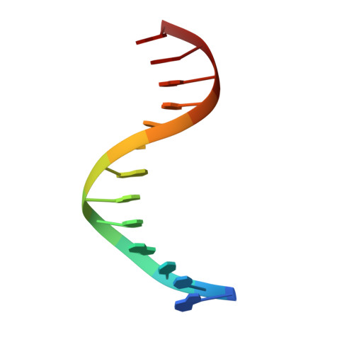
5'-D(*GP*TP*TP*AP*GP*GP*GP*TP*TP*AP*GP*GP*G)-3'A [auth B]13N/A
Sequence Annotations
Expand
  • Reference Sequence

Find similar nucleic acids by:  Sequence   |   3D Structure  

Entity ID: 2
MoleculeChains LengthOrganismImage
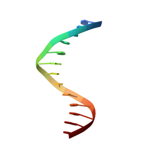
5'-D(*CP*CP*CP*TP*AP*AP*CP*CP*CP*TP*AP*AP*C)-3'B [auth C]13N/A
Sequence Annotations
Expand
  • Reference Sequence
Experimental Data & Validation

Experimental Data

  • Method: SOLUTION NMR
  • Conformers Calculated: 100 
  • Conformers Submitted: 20 
  • Selection Criteria: structures with the least restraint violations 

Structure Validation

View Full Validation Report


Entry History 

Revision History  (Full details and data files)

  • Version 1.0: 2002-04-17
    Type: Initial release
  • Version 1.1: 2008-04-27
    Changes: Version format compliance
  • Version 1.2: 2011-07-13
    Changes: Version format compliance
  • Version 1.3: 2022-02-23
    Changes: Database references, Derived calculations
  • Version 1.4: 2023-12-27
    Changes: Data collection