1ITB

TYPE-1 INTERLEUKIN-1 RECEPTOR COMPLEXED WITH INTERLEUKIN-1 BETA


Experimental Data Snapshot

  • Method: X-RAY DIFFRACTION
  • Resolution: 2.50 Å
  • R-Value Free: 0.325 
  • R-Value Work: 0.229 
  • R-Value Observed: 0.229 

wwPDB Validation   3D Report Full Report


This is version 1.2 of the entry. See complete history


Literature

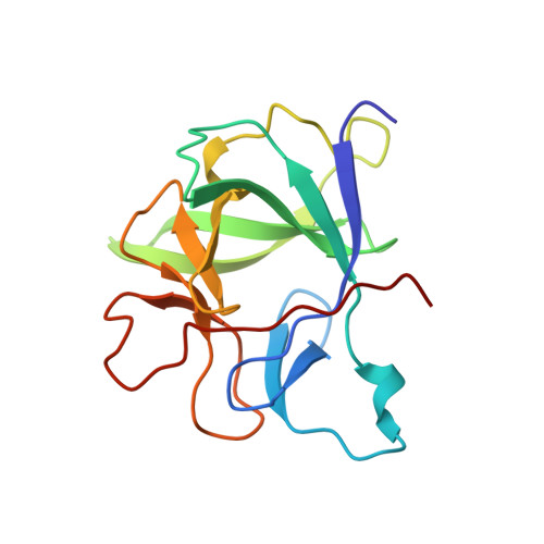
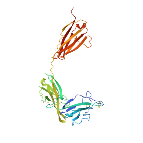
Crystal structure of the type-I interleukin-1 receptor complexed with interleukin-1beta.

Vigers, G.P.Anderson, L.J.Caffes, P.Brandhuber, B.J.

(1997) Nature 386: 190-194

  • DOI: https://doi.org/10.1038/386190a0
  • Primary Citation of Related Structures:  
    1ITB

  • PubMed Abstract: 
  • Interleukin-1 (IL-1) is an important mediator of inflammatory disease. The IL-1 family currently consists of two agonists, IL-1alpha and IL-1beta, and one antagonist, IL-1ra. Each of these molecules binds to the type I IL-1 receptor (IL1R). The binding of IL-1alpha or IL-1beta to IL1R is an early step in IL-1 signal transduction and blocking this interaction may therefore be a useful target for the development of new drugs ...

    Interleukin-1 (IL-1) is an important mediator of inflammatory disease. The IL-1 family currently consists of two agonists, IL-1alpha and IL-1beta, and one antagonist, IL-1ra. Each of these molecules binds to the type I IL-1 receptor (IL1R). The binding of IL-1alpha or IL-1beta to IL1R is an early step in IL-1 signal transduction and blocking this interaction may therefore be a useful target for the development of new drugs. Here we report the three-dimensional structure of IL-1beta bound to the extracellular domain of IL1R (s-IL1R) at 2.5 A resolution. IL-1beta binds to s-IL1R with a 1:1 stoichiometry. The crystal structure shows that s-IL1R consists of three immunoglobulin-like domains which wrap around IL-1beta in a manner distinct from the structures of previously described cytokine-receptor complexes. The two receptor-binding regions on IL-1beta identified by site-directed mutagenesis both contact the receptor: one binds to the first two domains of the receptor, while the other binds exclusively to the third domain.


    Organizational Affiliation

    Amgen Inc., Boulder, Colorado 80301, USA.


Macromolecules
Find similar proteins by:  (by identity cutoff)  |  3D Structure
Entity ID: 1
MoleculeChainsSequence LengthOrganismDetailsImage
INTERLEUKIN-1 BETA153Homo sapiensMutation(s): 0 
Gene Names: IL1BIL1F2
UniProt & NIH Common Fund Data Resources
Find proteins for P01584 (Homo sapiens)
Explore P01584 
Go to UniProtKB:  P01584
PHAROS:  P01584
Entity Groups  
Sequence Clusters30% Identity50% Identity70% Identity90% Identity95% Identity100% Identity
UniProt GroupP01584
Protein Feature View
Expand
  • Reference Sequence
Find similar proteins by:  (by identity cutoff)  |  3D Structure
Entity ID: 2
MoleculeChainsSequence LengthOrganismDetailsImage
TYPE 1 INTERLEUKIN-1 RECEPTOR315Homo sapiensMutation(s): 0 
Gene Names: IL1R1IL1RIL1RAIL1RT1
EC: 3.2.2.6
UniProt & NIH Common Fund Data Resources
Find proteins for P14778 (Homo sapiens)
Explore P14778 
Go to UniProtKB:  P14778
PHAROS:  P14778
Entity Groups  
Sequence Clusters30% Identity50% Identity70% Identity90% Identity95% Identity100% Identity
UniProt GroupP14778
Protein Feature View
Expand
  • Reference Sequence
Experimental Data & Validation

Experimental Data

  • Method: X-RAY DIFFRACTION
  • Resolution: 2.50 Å
  • R-Value Free: 0.325 
  • R-Value Work: 0.229 
  • R-Value Observed: 0.229 
  • Space Group: C 1 2 1
Unit Cell:
Length ( Å )Angle ( ˚ )
a = 146.952α = 90
b = 68.452β = 108.95
c = 65.867γ = 90
Software Package:
Software NamePurpose
DENZOdata reduction
SCALEPACKdata scaling
X-PLORmodel building
X-PLORrefinement
X-PLORphasing

Structure Validation

View Full Validation Report


Entry History 

Deposition Data

Revision History  (Full details and data files)

  • Version 1.0: 1998-02-04
    Type: Initial release
  • Version 1.1: 2008-03-24
    Changes: Version format compliance
  • Version 1.2: 2011-07-13
    Changes: Version format compliance