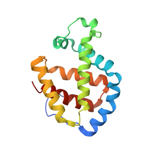
Crystal structures of deoxy- and carbonmonoxyhemoglobin F1 from the hagfish Eptatretus burgeri
Mito, M., Chong, K.T., Miyazaki, G., Adachi, S., Park, S.-Y., Tame, J.R., Morimoto, H.(2002) J Biological Chem 277: 21898-21905
- PubMed: 11923284
- DOI: https://doi.org/10.1074/jbc.M111492200
- Primary Citation of Related Structures:
1IT2, 1IT3 - PubMed Abstract:
Hagfish are extremely primitive jawless fish of disputed ancestry. Although generally classed with lampreys as cyclostomes ("round mouths"), it is clear that they diverged from them several hundred million years ago. The crystal structures of the deoxy and CO forms of hemoglobin from a hagfish (Eptatretus burgeri) have been solved at 1.6 and 2.1 A, respectively. The deoxy crystal contains one dimer and two monomers in a unit cell, with the dimer being similar to that found in lamprey deoxy-Hb, but with a larger interface and different relative orientation of the partner chains. Ile(E11) and Gln(E7) obstruct ligand binding in the deoxy form and make room for ligands in the CO form, but no interaction path between the two hemes could be identified. The BGH core structure, which forms the alpha1beta1 interface of all vertebrate alpha2beta2 tetrameric Hbs, is conserved in hagfish and lamprey Hbs. It was shown previously that human and cartilaginous fish Hbs have independently evolved stereochemical mechanisms other than the movement of the proximal histidine to regulate ligand binding at the hemes. Our results therefore suggest that the formation of the alpha2beta2 tetramer using the BGH core and the mechanism of quaternary structure change evolved between the branching points of hagfish and lampreys from other vertebrates.
- Division of Biophysical Engineering, Graduate School of Engineering Science, Osaka University, Toyonaka 560-8531 Osaka, Japan.
Organizational Affiliation:


