1ISN

Crystal structure of merlin FERM domain


Experimental Data Snapshot

  • Method: X-RAY DIFFRACTION
  • Resolution: 2.90 Å
  • R-Value Free: 0.284 
  • R-Value Work: 0.229 
  • R-Value Observed: 0.235 

wwPDB Validation   3D Report Full Report


This is version 1.3 of the entry. See complete history


Literature

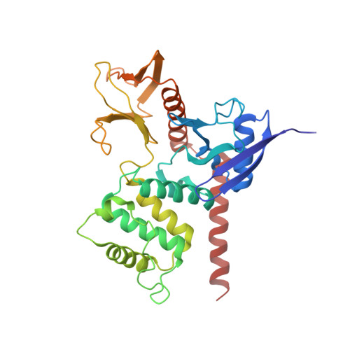
Structural basis for neurofibromatosis type 2. Crystal structure of the merlin FERM domain.

Shimizu, T.Seto, A.Maita, N.Hamada, K.Tsukita, S.Tsukita, S.Hakoshima, T.

(2002) J Biol Chem 277: 10332-10336

  • DOI: https://doi.org/10.1074/jbc.M109979200
  • Primary Citation of Related Structures:  
    1ISN

  • PubMed Abstract: 

    Neurofibromatosis type 2 (NF2) is a dominantly inherited disease associated with the central nervous system. The NF2 gene product merlin is a tumor suppressor, and its mutation or inactivation causes this disease. We report here the crystal structure of the merlin FERM domain containing a 22-residue alpha-helical segment. The structure reveals that the merlin FERM domain consists of three subdomains displaying notable features of the electrostatic surface potentials, although the overall surface potentials similar to those of ezrin/radixin/moesin (ERM) proteins indicate electrostatic membrane association. The structure also is consistent with inactivation mechanisms caused by the pathogenic mutations associated with NF2.


  • Organizational Affiliation

    Structural Biology Laboratory, Nara Institute of Science and Technology and CREST, Japan.


Macromolecules
Find similar proteins by:  (by identity cutoff)  |  3D Structure
Entity ID: 1
MoleculeChains Sequence LengthOrganismDetailsImage
merlin323Mus musculusMutation(s): 0 
Gene Names: neurofibromatosis type 2
UniProt
Find proteins for P46662 (Mus musculus)
Explore P46662 
Go to UniProtKB:  P46662
Entity Groups  
Sequence Clusters30% Identity50% Identity70% Identity90% Identity95% Identity100% Identity
UniProt GroupP46662
Sequence Annotations
Expand
  • Reference Sequence
Experimental Data & Validation

Experimental Data

  • Method: X-RAY DIFFRACTION
  • Resolution: 2.90 Å
  • R-Value Free: 0.284 
  • R-Value Work: 0.229 
  • R-Value Observed: 0.235 
  • Space Group: P 43 21 2
Unit Cell:
Length ( Å )Angle ( ˚ )
a = 67.408α = 90
b = 67.408β = 90
c = 179.137γ = 90
Software Package:
Software NamePurpose
AMoREphasing
CNSrefinement
DPSdata reduction
MOSFLMdata reduction

Structure Validation

View Full Validation Report


Entry History 

Deposition Data

Revision History  (Full details and data files)

  • Version 1.0: 2002-04-03
    Type: Initial release
  • Version 1.1: 2008-04-27
    Changes: Version format compliance
  • Version 1.2: 2011-07-13
    Changes: Version format compliance
  • Version 1.3: 2023-10-25
    Changes: Data collection, Database references, Refinement description