1IMJ | pdb_00001imj

CRYSTAL STRUCTURE OF THE HUMAN CCG1/TAFII250-INTERACTING FACTOR B (CIB)


Experimental Data Snapshot

  • Method: X-RAY DIFFRACTION
  • Resolution: 2.20 Å
  • R-Value Free: 
    0.235 (Depositor) 
  • R-Value Work: 
    0.189 (Depositor) 
  • R-Value Observed: 
    0.189 (Depositor) 

wwPDB Validation 3D Report Full Report

Validation slider image for 1IMJ

This is version 1.3 of the entry. See complete history

Literature

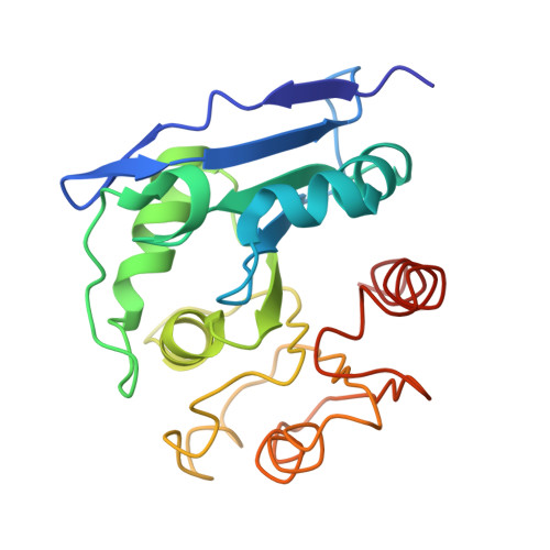
The crystal structure of CCG1/TAF(II)250-interacting factor B (CIB)

Padmanabhan, B.Kuzuhara, T.Adachi, N.Horikoshi, M.

(2004) J Biological Chem 279: 9615-9624

  • DOI: https://doi.org/10.1074/jbc.M312165200
  • Primary Citation Related Structures: 
    1IMJ

  • PubMed Abstract: 

    The general transcription initiation factor TFIID and its interactors play critical roles in regulating the transcription from both naked and chromatin DNA. We have isolated a novel TFIID interactor that we denoted as CCG1/TAF(II)250-interacting factor B (CIB). We show here that CIB activates transcription. To further understand the function of this protein, we determined its crystal structure at 2.2-Angstroms resolution. The tertiary structure of CIB reveals an alpha/beta-hydrolase fold that resembles structures in the prokaryotic alpha/beta-hydrolase family proteins. It is not similar in structure or primary sequence to any eukaryotic transcription or chromatin factors that have been reported to date. CIB possesses a conserved catalytic triad that is found in other alpha/beta-hydrolases, and our in vitro studies confirmed that it bears hydrolase activity. However, CIB differs from other alpha/beta-hydrolases in that it lacks a binding site excursion, which facilitates the substrate selectivity of the other alpha/beta-hydrolases. Further functional characterization of CIB based on its tertiary structure and through biochemical studies may provide novel insights into the mechanisms that regulate eukaryotic transcription.


  • Organizational Affiliation
    • Horikoshi Gene Selector Project, Exploratory Research for Advanced Technology, Japan Science and Technology Corporation, 5-9-6 Tokodai, Tsukuba, Ibaraki 300-2635, Japan.

Macromolecule Content 

  • Total Structure Weight: 22.46 kDa 
  • Atom Count: 1,756 
  • Modeled Residue Count: 208 
  • Deposited Residue Count: 210 
  • Unique protein chains: 1

Macromolecules

Find similar proteins by:|  3D Structure
Entity ID: 1
MoleculeChains  Sequence LengthOrganismDetailsImage
CCG1-INTERACTING FACTOR B210Homo sapiensMutation(s): 0 
EC: 2.3.1
UniProt & NIH Common Fund Data Resources
Find proteins for Q96IU4 (Homo sapiens)
Explore Q96IU4 
Go to UniProtKB:  Q96IU4
PHAROS:  Q96IU4
GTEx:  ENSG00000114779 
Entity Groups
Sequence Clusters30% Identity50% Identity70% Identity90% Identity95% Identity100% Identity
UniProt GroupQ96IU4
Sequence Annotations
Expand
Reference Sequence

Small Molecules

Ligands 1 Unique
IDChains Name / Formula / InChI Key2D Diagram3D Interactions
SO4

Query on SO4


Download:Ideal Coordinates CCD File
B [auth A]SULFATE ION
O4 S
QAOWNCQODCNURD-UHFFFAOYSA-L

Experimental Data & Validation

Experimental Data

  • Method: X-RAY DIFFRACTION
  • Resolution: 2.20 Å
  • R-Value Free:  0.235 (Depositor) 
  • R-Value Work:  0.189 (Depositor) 
  • R-Value Observed: 0.189 (Depositor) 
Space Group: P 21 21 2
Unit Cell:
Length ( Å )Angle ( ˚ )
a = 110.704α = 90
b = 44.445β = 90
c = 43.601γ = 90
Software Package:
Software NamePurpose
SCALEPACKdata scaling
SOLVEphasing
CNSrefinement

Structure Validation

View Full Validation Report


Entry History 

Deposition Data

Revision History  (Full details and data files)

  • Version 1.0: 2002-05-11
    Type: Initial release
  • Version 1.1: 2008-04-27
    Changes: Version format compliance
  • Version 1.2: 2011-07-13
    Changes: Version format compliance
  • Version 1.3: 2024-03-13
    Changes: Data collection, Database references, Derived calculations