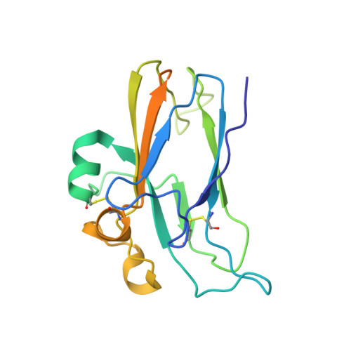
1IKO

CRYSTAL STRUCTURE OF THE MURINE EPHRIN-B2 ECTODOMAIN


Experimental Data Snapshot

  • Method: X-RAY DIFFRACTION
  • Resolution: 1.92 Å
  • R-Value Free: 0.249 
  • R-Value Work: 0.216 
  • R-Value Observed: 0.216 

wwPDB Validation   3D Report Full Report


This is version 2.1 of the entry. See complete history


Literature

Crystal structure of an ephrin ectodomain.

Toth, J.Cutforth, T.Gelinas, A.D.Bethoney, K.A.Bard, J.Harrison, C.J.

(2001) Dev Cell 1: 83-92

  • DOI: https://doi.org/10.1016/s1534-5807(01)00002-8
  • Primary Citation of Related Structures:  
    1IKO

  • PubMed Abstract: 

    Eph receptor tyrosine kinases and their membrane-associated ligands, the ephrins, are essential regulators of axon guidance, cell migration, segmentation, and angiogenesis. There are two classes of vertebrate ephrin ligands which have distinct binding specificities for their cognate receptors. Multimerization of the ligands is required for receptor activation, and ephrin ligands themselves signal intracellularly upon binding Eph receptors. We have determined the structure of the extracellular domain of mouse ephrin-B2. The ephrin ectodomain is an eight-stranded beta barrel with topological similarity to plant nodulins and phytocyanins. Based on the structure, we have identified potential surface determinants of Eph/ephrin binding specificity and a ligand dimerization region. The high sequence similarity among ephrin ectodomains indicates that all ephrins may be modeled upon the ephrin-B2 structure presented here.


  • Organizational Affiliation

    Boston Biomedical Research Institute, Watertown, Massachusetts 02472, USA.


Macromolecules
Find similar proteins by:  (by identity cutoff)  |  3D Structure
Entity ID: 1
MoleculeChains Sequence LengthOrganismDetailsImage
EPHRIN-B2A [auth P]178Mus musculusMutation(s): 0 
Gene Names: ephrin-B2
UniProt
Find proteins for P52800 (Mus musculus)
Explore P52800 
Go to UniProtKB:  P52800
Entity Groups  
Sequence Clusters30% Identity50% Identity70% Identity90% Identity95% Identity100% Identity
UniProt GroupP52800
Glycosylation
Glycosylation Sites: 1Go to GlyGen: P52800-1
Sequence Annotations
Expand
  • Reference Sequence
Oligosaccharides

Help

Entity ID: 2
MoleculeChains Length2D Diagram Glycosylation3D Interactions
alpha-D-mannopyranose-(1-4)-2-acetamido-2-deoxy-beta-D-glucopyranose-(1-4)-2-acetamido-2-deoxy-beta-D-glucopyranoseB [auth A]3N-Glycosylation
Glycosylation Resources
GlyTouCan:  G62182OO
GlyCosmos:  G62182OO
GlyGen:  G62182OO
Experimental Data & Validation

Experimental Data

  • Method: X-RAY DIFFRACTION
  • Resolution: 1.92 Å
  • R-Value Free: 0.249 
  • R-Value Work: 0.216 
  • R-Value Observed: 0.216 
  • Space Group: P 65 2 2
Unit Cell:
Length ( Å )Angle ( ˚ )
a = 59.283α = 90
b = 59.283β = 90
c = 168.868γ = 120
Software Package:
Software NamePurpose
HKL-2000data collection
HKL-2000data reduction
SHARPphasing
CNSrefinement
HKL-2000data scaling

Structure Validation

View Full Validation Report


Entry History 

Deposition Data

Revision History  (Full details and data files)

  • Version 1.0: 2002-05-03
    Type: Initial release
  • Version 1.1: 2008-04-27
    Changes: Version format compliance
  • Version 1.2: 2011-07-13
    Changes: Non-polymer description, Version format compliance
  • Version 2.0: 2020-07-29
    Type: Remediation
    Reason: Carbohydrate remediation
    Changes: Advisory, Atomic model, Data collection, Derived calculations, Structure summary
  • Version 2.1: 2024-11-06
    Changes: Data collection, Database references, Structure summary