1IJW

Testing the Water-Mediated Hin Recombinase DNA Recognition by Systematic Mutations.


Experimental Data Snapshot

  • Method: X-RAY DIFFRACTION
  • Resolution: 2.40 Å
  • R-Value Free: 0.278 
  • R-Value Work: 0.223 
  • R-Value Observed: 0.223 

Starting Model: experimental
View more details

wwPDB Validation   3D Report Full Report


This is version 1.4 of the entry. See complete history


Literature

Testing water-mediated DNA recognition by the Hin recombinase.

Chiu, T.K.Sohn, C.Dickerson, R.E.Johnson, R.C.

(2002) EMBO J 21: 801-814

  • DOI: https://doi.org/10.1093/emboj/21.4.801
  • Primary Citation of Related Structures:  
    1IJW, 1JJ6, 1JJ8, 1JKO, 1JKP, 1JKQ, 1JKR

  • PubMed Abstract: 

    The Hin recombinase specifically recognizes its DNA-binding site by means of both major and minor groove interactions. A previous X-ray structure, together with new structures of the Hin DNA-binding domain bound to a recombination half-site that were solved as part of the present study, have revealed that two ordered water molecules are present within the major groove interface. In this report, we test the importance of these waters directly by X-ray crystal structure analysis of complexes with four mutant DNA sequences. These structures, combined with their Hin-binding properties, provide strong support for the critical importance of one of the intermediate waters. A lesser but demonstrable role is ascribed to the second water molecule. The mutant structures also illustrate the prominent roles of thymine methyls both in stabilizing intermediate waters and in interfering with water or amino acid side chain interactions with DNA.


  • Organizational Affiliation

    Department of Chemistry and Biochemistry, University of California at Los Angeles, Los Angeles, CA 90095, USA.


Macromolecules

Find similar proteins by:  (by identity cutoff)  |  3D Structure
Entity ID: 3
MoleculeChains Sequence LengthOrganismDetailsImage
DNA-INVERTASE HIN52N/AMutation(s): 0 
UniProt
Find proteins for P03013 (Salmonella typhimurium (strain LT2 / SGSC1412 / ATCC 700720))
Explore P03013 
Go to UniProtKB:  P03013
Entity Groups  
Sequence Clusters30% Identity50% Identity70% Identity90% Identity95% Identity100% Identity
UniProt GroupP03013
Sequence Annotations
Expand
  • Reference Sequence

Find similar nucleic acids by:  Sequence   |   3D Structure  

Entity ID: 1
MoleculeChains LengthOrganismImage
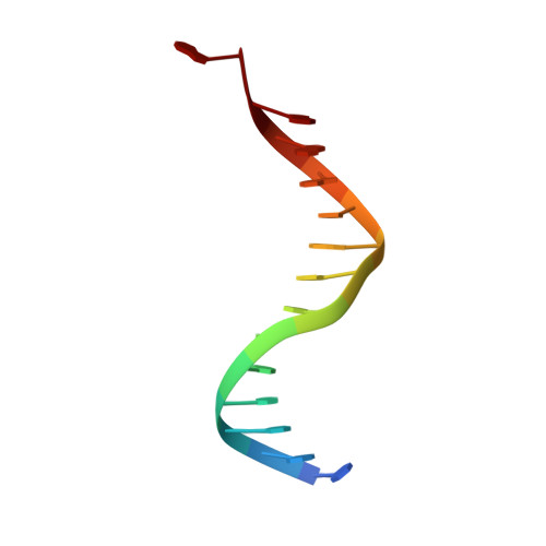
5'-D(*TP*GP*TP*TP*TP*TP*TP*GP*AP*TP*AP*AP*GP*A)-3'14N/A
Sequence Annotations
Expand
  • Reference Sequence

Find similar nucleic acids by:  Sequence   |   3D Structure  

Entity ID: 2
MoleculeChains LengthOrganismImage
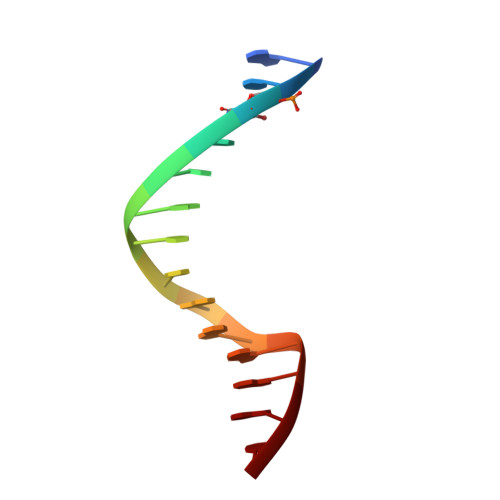
5'-D(*AP*TP*(CBR)P*TP*TP*AP*TP*CP*AP*AP*AP*AP*AP*C)-3'14N/A
Sequence Annotations
Expand
  • Reference Sequence
Small Molecules
Ligands 1 Unique
IDChains Name / Formula / InChI Key2D Diagram3D Interactions
TRS
Query on TRS

Download Ideal Coordinates CCD File 
D [auth B],
E [auth C]
2-AMINO-2-HYDROXYMETHYL-PROPANE-1,3-DIOL
C4 H12 N O3
LENZDBCJOHFCAS-UHFFFAOYSA-O
Binding Affinity Annotations 
IDSourceBinding Affinity
DNA PDBBind:  1IJW Kd: 3.8 (nM) from 1 assay(s)
Experimental Data & Validation

Experimental Data

  • Method: X-RAY DIFFRACTION
  • Resolution: 2.40 Å
  • R-Value Free: 0.278 
  • R-Value Work: 0.223 
  • R-Value Observed: 0.223 
  • Space Group: C 2 2 21
Unit Cell:
Length ( Å )Angle ( ˚ )
a = 85.749α = 90
b = 81.136β = 90
c = 44.596γ = 90
Software Package:
Software NamePurpose
SOLVEphasing
CNSrefinement
DENZOdata reduction
SCALEPACKdata scaling

Structure Validation

View Full Validation Report


Entry History 

Deposition Data

Revision History  (Full details and data files)

  • Version 1.0: 2002-02-22
    Type: Initial release
  • Version 1.1: 2008-04-27
    Changes: Version format compliance
  • Version 1.2: 2011-07-13
    Changes: Version format compliance
  • Version 1.3: 2024-02-07
    Changes: Data collection, Database references, Derived calculations
  • Version 1.4: 2024-04-03
    Changes: Refinement description