1IHJ

Crystal Structure of the N-terminal PDZ domain of InaD in complex with a NorpA C-terminal peptide


Experimental Data Snapshot

  • Method: X-RAY DIFFRACTION
  • Resolution: 1.80 Å
  • R-Value Free: 0.240 
  • R-Value Work: 0.220 
  • R-Value Observed: 0.220 

wwPDB Validation   3D Report Full Report


This is version 1.3 of the entry. See complete history


Literature

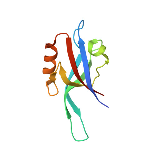
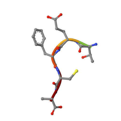
Functional relevance of the disulfide-linked complex of the N-terminal PDZ domain of InaD with NorpA.

Kimple, M.E.Siderovski, D.P.Sondek, J.

(2001) EMBO J 20: 4414-4422

  • DOI: https://doi.org/10.1093/emboj/20.16.4414
  • Primary Citation of Related Structures:  
    1IHJ

  • PubMed Abstract: 
  • In Drosophila, phototransduction is mediated by G(q)-activation of phospholipase C and is a well studied model system for understanding the kinetics of signal initiation, propagation and termination controlled by G proteins. The proper intracellular targeting and spatial arrangement of most proteins involved in fly phototransduction require the multi-domain scaffolding protein InaD, composed almost entirely of five PDZ domains, which independently bind various proteins including NorpA, the relevant phospho lipase C-beta isozyme ...

    In Drosophila, phototransduction is mediated by G(q)-activation of phospholipase C and is a well studied model system for understanding the kinetics of signal initiation, propagation and termination controlled by G proteins. The proper intracellular targeting and spatial arrangement of most proteins involved in fly phototransduction require the multi-domain scaffolding protein InaD, composed almost entirely of five PDZ domains, which independently bind various proteins including NorpA, the relevant phospho lipase C-beta isozyme. We have determined the crystal structure of the N-terminal PDZ domain of InaD bound to a peptide corresponding to the C-terminus of NorpA to 1.8 A resolution. The structure highlights an intermolecular disulfide bond necessary for high affinity interaction as determined by both in vitro and in vivo studies. Since other proteins also possess similar, cysteine-containing consensus sequences for binding PDZ domains, this disulfide-mediated 'dock-and-lock' interaction of PDZ domains with their ligands may be a relatively ubiquitous mode of coordinating signaling pathways.


    Organizational Affiliation

    Department of Biochemistry and Biophysics, The University of North Carolina at Chapel Hill, Chapel Hill, NC 27599, USA.


Macromolecules
Find similar proteins by:  (by identity cutoff)  |  3D Structure
Entity ID: 1
MoleculeChainsSequence LengthOrganismDetailsImage
InaD
A, B
98Drosophila melanogasterMutation(s): 0 
Gene Names: inaDCG3504
UniProt
Find proteins for Q24008 (Drosophila melanogaster)
Explore Q24008 
Go to UniProtKB:  Q24008
Entity Groups  
Sequence Clusters30% Identity50% Identity70% Identity90% Identity95% Identity100% Identity
UniProt GroupQ24008
Protein Feature View
Expand
  • Reference Sequence

Find similar proteins by:  Sequence   |   3D Structure  

Entity ID: 2
MoleculeChainsSequence LengthOrganismDetailsImage
phospholipase C
C, D
7N/AMutation(s): 0 
EC: 3.1.4.3
Protein Feature View
Expand
  • Reference Sequence
Experimental Data & Validation

Experimental Data

  • Method: X-RAY DIFFRACTION
  • Resolution: 1.80 Å
  • R-Value Free: 0.240 
  • R-Value Work: 0.220 
  • R-Value Observed: 0.220 
  • Space Group: P 1
Unit Cell:
Length ( Å )Angle ( ˚ )
a = 42.17α = 106.55
b = 44.15β = 100.61
c = 44.46γ = 118.25
Software Package:
Software NamePurpose
DENZOdata reduction
SCALEPACKdata scaling
AMoREphasing
CNSrefinement

Structure Validation

View Full Validation Report


Entry History 

Deposition Data

Revision History  (Full details and data files)

  • Version 1.0: 2001-08-22
    Type: Initial release
  • Version 1.1: 2008-04-27
    Changes: Version format compliance
  • Version 1.2: 2011-07-13
    Changes: Version format compliance
  • Version 1.3: 2023-08-16
    Changes: Data collection, Database references, Refinement description