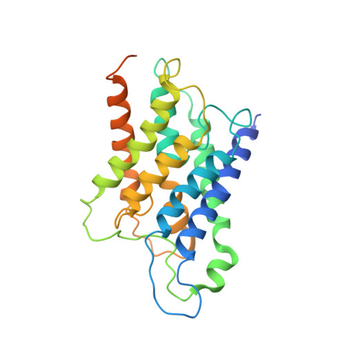
1IH5

CRYSTAL STRUCTURE OF AQUAPORIN-1


Experimental Data Snapshot

  • Method: ELECTRON CRYSTALLOGRAPHY
  • Resolution: 3.70 Å
  • R-Value Free: 0.458 
  • R-Value Work: 0.361 
  • R-Value Observed: 0.361 

wwPDB Validation   3D Report Full Report


This is version 1.3 of the entry. See complete history


Literature

Visualization of a water-selective pore by electron crystallography in vitreous ice.

Ren, G.Reddy, V.S.Cheng, A.Melnyk, P.Mitra, A.K.

(2001) Proc Natl Acad Sci U S A 98: 1398-1403

  • DOI: https://doi.org/10.1073/pnas.98.4.1398
  • Primary Citation of Related Structures:  
    1IH5

  • PubMed Abstract: 

    The water-selective pathway through the aquaporin-1 membrane channel has been visualized by fitting an atomic model to a 3.7-A resolution three-dimensional density map. This map was determined by analyzing images and electron diffraction patterns of lipid-reconstituted two-dimensional crystals of aquaporin-1 preserved in vitrified buffer in the absence of any additive. The aqueous pathway is characterized by a size-selective pore that is approximately 4.0 +/- 0.5A in diameter, spans a length of approximately 18A, and bends by approximately 25 degrees as it traverses the bilayer. This narrow pore is connected by wide, funnel-shaped openings at the extracellular and cytoplasmic faces. The size-selective pore is outlined mostly by hydrophobic residues, resulting in a relatively inert pathway conducive to diffusion-limited water flow. The apex of the curved pore is close to the locations of the in-plane pseudo-2-fold symmetry axis that relates the N- and C-terminal halves and the conserved, functionally important N76 and N192 residues.


  • Organizational Affiliation

    Department of Cell Biology, The Scripps Research Institute, 10550 North Torrey Pines Road, La Jolla, CA 92037, USA.


Macromolecules
Find similar proteins by:  (by identity cutoff)  |  3D Structure
Entity ID: 1
MoleculeChains Sequence LengthOrganismDetailsImage
AQUAPORIN-1269Homo sapiensMutation(s): 0 
Membrane Entity: Yes 
UniProt & NIH Common Fund Data Resources
Find proteins for P29972 (Homo sapiens)
Explore P29972 
Go to UniProtKB:  P29972
PHAROS:  P29972
GTEx:  ENSG00000240583 
Entity Groups  
Sequence Clusters30% Identity50% Identity70% Identity90% Identity95% Identity100% Identity
UniProt GroupP29972
Sequence Annotations
Expand
  • Reference Sequence
Experimental Data & Validation

Experimental Data

  • Method: ELECTRON CRYSTALLOGRAPHY
  • Resolution: 3.70 Å
  • R-Value Free: 0.458 
  • R-Value Work: 0.361 
  • R-Value Observed: 0.361 
  • Space Group: P 4 21 2
Unit Cell:
Length ( Å )Angle ( ˚ )
a = 99.58α = 90
b = 99.58β = 90
c = 100γ = 90
Software Package:
Software NamePurpose
X-PLORrefinement

Structure Validation

View Full Validation Report


Entry History 

Deposition Data

Revision History  (Full details and data files)

  • Version 1.0: 2001-04-25
    Type: Initial release
  • Version 1.1: 2008-04-27
    Changes: Version format compliance
  • Version 1.2: 2011-07-13
    Changes: Derived calculations, Version format compliance
  • Version 1.3: 2024-02-07
    Changes: Author supporting evidence, Data collection, Database references