Structural basis for the negative allostery between Ca(2+)- and Mg(2+)-binding in the intracellular Ca(2+)-receptor calbindin D9k.
Andersson, M., Malmendal, A., Linse, S., Ivarsson, I., Forsen, S., Svensson, L.A.(1997) Protein Sci 6: 1139-1147
- PubMed: 9194174 Search on PubMedSearch on PubMed Central
- DOI: https://doi.org/10.1002/pro.5560060602
- Primary Citation Related Structures:
1IG5, 1IGV - PubMed Abstract:
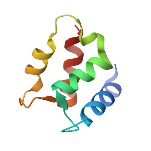
The three-dimensional structures of the magnesium- and manganese-bound forms of calbindin D9k were determined to 1.6 A and 1.9 A resolution, respectively, using X-ray crystallography. These two structures are nearly identical but deviate significantly from both the calcium bound form and the metal ion-free (apo) form. The largest structural differences are seen in the C-terminal EF-hand, and involve changes in both metal ion coordination and helix packing. The N-terminal calcium binding site is not occupied by any metal ion in the magnesium and manganese structures, and shows little structural deviation from the apo and calcium bound forms. 1H-NMR and UV spectroscopic studies at physiological ion concentrations show that the C-terminal site of the protein is significantly populated by magnesium at resting cell calcium levels, and that there is a negative allosteric interaction between magnesium and calcium binding. Calcium binding was found to occur with positive cooperativity at physiological magnesium concentration.
- Department of Molecular Biophysics, Lund University, Sweden.
Organizational Affiliation: