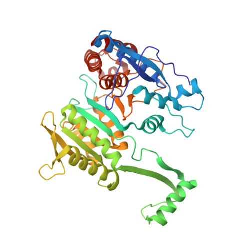
1IDC

ISOCITRATE DEHYDROGENASE FROM E.COLI (MUTANT K230M), STEADY-STATE INTERMEDIATE COMPLEX DETERMINED BY LAUE CRYSTALLOGRAPHY


Experimental Data Snapshot

  • Method: X-RAY DIFFRACTION
  • Resolution: 2.50 Å
  • R-Value Work: 0.169 
  • R-Value Observed: 0.169 

wwPDB Validation   3D Report Full Report


This is version 1.4 of the entry. See complete history


Literature

Mutagenesis and Laue structures of enzyme intermediates: isocitrate dehydrogenase.

Bolduc, J.M.Dyer, D.H.Scott, W.G.Singer, P.Sweet, R.M.Koshland Jr., D.E.Stoddard, B.L.

(1995) Science 268: 1312-1318

  • DOI: https://doi.org/10.1126/science.7761851
  • Primary Citation of Related Structures:  
    1IDC, 1IDD, 1IDE, 1IDF

  • PubMed Abstract: 

    Site-directed mutagenesis and Laue diffraction data to 2.5 A resolution were used to solve the structures of two sequential intermediates formed during the catalytic actions of isocitrate dehydrogenase. Both intermediates are distinct from the enzyme-substrate and enzyme-product complexes. Mutation of key catalytic residues changed the rate determining steps so that protein and substrate intermediates within the overall reaction pathway could be visualized.


  • Organizational Affiliation

    Fred Hutchinson Cancer Research Center, Program in Structural Biology, Seattle, WA 98104, USA.


Macromolecules
Find similar proteins by:  (by identity cutoff)  |  3D Structure
Entity ID: 1
MoleculeChains Sequence LengthOrganismDetailsImage
ISOCITRATE DEHYDROGENASE416Escherichia coliMutation(s): 1 
Gene Names: ICD
EC: 1.1.1.42
UniProt
Find proteins for P08200 (Escherichia coli (strain K12))
Explore P08200 
Go to UniProtKB:  P08200
Entity Groups  
Sequence Clusters30% Identity50% Identity70% Identity90% Identity95% Identity100% Identity
UniProt GroupP08200
Sequence Annotations
Expand
  • Reference Sequence
Small Molecules
Ligands 2 Unique
IDChains Name / Formula / InChI Key2D Diagram3D Interactions
OXS
Query on OXS

Download Ideal Coordinates CCD File 
C [auth A]2-OXALOSUCCINIC ACID
C6 H6 O7
UFSCUAXLTRFIDC-REOHCLBHSA-N
MG
Query on MG

Download Ideal Coordinates CCD File 
B [auth A]MAGNESIUM ION
Mg
JLVVSXFLKOJNIY-UHFFFAOYSA-N
Experimental Data & Validation

Experimental Data

  • Method: X-RAY DIFFRACTION
  • Resolution: 2.50 Å
  • R-Value Work: 0.169 
  • R-Value Observed: 0.169 
  • Space Group: P 43 21 2
Unit Cell:
Length ( Å )Angle ( ˚ )
a = 105.1α = 90
b = 105.1β = 90
c = 150.3γ = 90
Software Package:
Software NamePurpose
X-PLORmodel building
X-PLORrefinement
OXFORDdata reduction
X-PLORphasing

Structure Validation

View Full Validation Report


Entry History 

Deposition Data

Revision History  (Full details and data files)

  • Version 1.0: 1996-03-08
    Type: Initial release
  • Version 1.1: 2008-03-03
    Changes: Version format compliance
  • Version 1.2: 2011-07-13
    Changes: Derived calculations, Version format compliance
  • Version 1.3: 2017-11-29
    Changes: Advisory, Derived calculations, Other
  • Version 1.4: 2021-11-03
    Changes: Advisory, Database references, Derived calculations