1IBC

CRYSTAL STRUCTURE OF INHIBITED INTERLEUKIN-1BETA CONVERTING ENZYME


Experimental Data Snapshot

  • Method: X-RAY DIFFRACTION
  • Resolution: 2.73 Å
  • R-Value Free: 0.285 
  • R-Value Work: 0.173 
  • R-Value Observed: 0.173 

wwPDB Validation   3D Report Full Report


This is version 1.6 of the entry. See complete history


Literature

A combinatorial approach for determining protease specificities: application to interleukin-1beta converting enzyme (ICE).

Rano, T.A.Timkey, T.Peterson, E.P.Rotonda, J.Nicholson, D.W.Becker, J.W.Chapman, K.T.Thornberry, N.A.

(1997) Chem Biol 4: 149-155

  • DOI: https://doi.org/10.1016/s1074-5521(97)90258-1
  • Primary Citation of Related Structures:  
    1IBC

  • PubMed Abstract: 

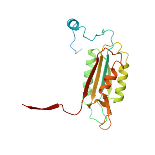
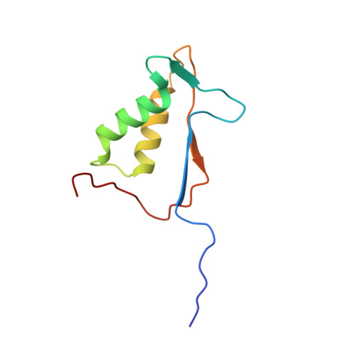
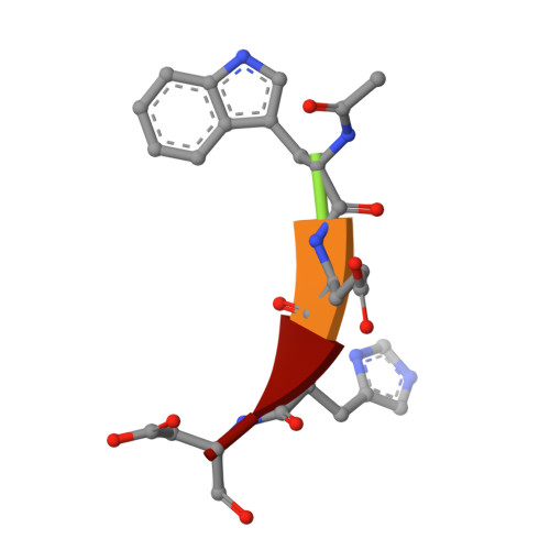
    Interleukin-1beta converting enzyme (ICE/caspase-1) is the protease responsible for interleukin-1beta (IL-1beta) production in monocytes. It was the first member of a new cysteine protease family to be identified. Members of this family have functions in both inflammation and apoptosis. A novel method for identifying protease specificity, employing a positional-scanning substrate library, was used to determine the amino-acid preferences of ICE. Using this method, the complete specificity of a protease can be mapped in the time required to perform one assay. The results indicate that the optimal tetrapeptide recognition sequence for ICE is WEHD, not YVAD, as previously believed, and this led to the synthesis of an unusually potent aldehyde inhibitor, Ac-WEHD-CHO (Ki = 56 pM). The structural basis for this potent inhibition was determined by X-ray crystallography. The results presented in this study establish a positional-scanning library as a powerful tool for rapidly and accurately assessing protease specificity. The preferred sequence for ICE (WEHD) differs significantly from that found in human pro-interleukin-1beta (YVHD), which suggests that this protease may have additional endogenous substrates, consistent with evidence linking it to apoptosis and IL-1alpha production.


  • Organizational Affiliation

    Department of Molecular Design and Diversity, Merck Research Laboratories, R123-232, PO Box 2000, Rahway, New Jersey 07065, USA.


Macromolecules
Find similar proteins by:  (by identity cutoff)  |  3D Structure
Entity ID: 1
MoleculeChains Sequence LengthOrganismDetailsImage
INTERLEUKIN-1BETA CONVERTING ENZYME194Homo sapiensMutation(s): 0 
EC: 3.4.22.36
UniProt & NIH Common Fund Data Resources
Find proteins for P29466 (Homo sapiens)
Explore P29466 
Go to UniProtKB:  P29466
PHAROS:  P29466
GTEx:  ENSG00000137752 
Entity Groups  
Sequence Clusters30% Identity50% Identity70% Identity90% Identity95% Identity100% Identity
UniProt GroupP29466
Sequence Annotations
Expand
  • Reference Sequence
Find similar proteins by:  (by identity cutoff)  |  3D Structure
Entity ID: 2
MoleculeChains Sequence LengthOrganismDetailsImage
INTERLEUKIN-1BETA CONVERTING ENZYME88Homo sapiensMutation(s): 1 
EC: 3.4.22.36
UniProt & NIH Common Fund Data Resources
Find proteins for P29466 (Homo sapiens)
Explore P29466 
Go to UniProtKB:  P29466
PHAROS:  P29466
GTEx:  ENSG00000137752 
Entity Groups  
Sequence Clusters30% Identity50% Identity70% Identity90% Identity95% Identity100% Identity
UniProt GroupP29466
Sequence Annotations
Expand
  • Reference Sequence

Find similar proteins by:  Sequence   |   3D Structure  

Entity ID: 3
MoleculeChains Sequence LengthOrganismDetailsImage
PEPTIDE ACE-TRP-GLU-HIS-ASA5N/AMutation(s): 0 
Sequence Annotations
Expand
  • Reference Sequence
Small Molecules
Modified Residues  1 Unique
IDChains TypeFormula2D DiagramParent
ASA
Query on ASA
C
L-PEPTIDE LINKINGC4 H7 N O3ASP
Experimental Data & Validation

Experimental Data

  • Method: X-RAY DIFFRACTION
  • Resolution: 2.73 Å
  • R-Value Free: 0.285 
  • R-Value Work: 0.173 
  • R-Value Observed: 0.173 
  • Space Group: P 43 21 2
Unit Cell:
Length ( Å )Angle ( ˚ )
a = 64.61α = 90
b = 64.61β = 90
c = 160.1γ = 90
Software Package:
Software NamePurpose
X-PLORmodel building
X-PLORrefinement
SAINTdata reduction
SAINTdata scaling
X-PLORphasing

Structure Validation

View Full Validation Report


Entry History 

Deposition Data

Revision History  (Full details and data files)

  • Version 1.0: 1998-02-11
    Type: Initial release
  • Version 1.1: 2008-03-24
    Changes: Version format compliance
  • Version 1.2: 2011-07-13
    Changes: Atomic model, Database references, Derived calculations, Non-polymer description, Structure summary, Version format compliance
  • Version 1.3: 2011-11-16
    Changes: Atomic model
  • Version 1.4: 2017-11-29
    Changes: Derived calculations, Other
  • Version 1.5: 2021-11-03
    Changes: Database references, Derived calculations
  • Version 1.6: 2024-04-03
    Changes: Data collection, Refinement description