1I4X

STAPHYLOCOCCAL ENTEROTOXIN C2, MONOCLINIC FORM CRYSTALLIZED AT PH 8.0


Experimental Data Snapshot

  • Method: X-RAY DIFFRACTION
  • Resolution: 2.40 Å
  • R-Value Work: 0.207 
  • R-Value Observed: 0.207 

wwPDB Validation   3D Report Full Report


This is version 1.3 of the entry. See complete history


Literature

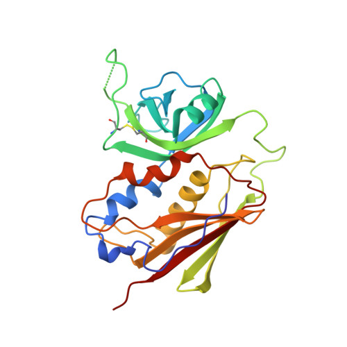
Structure of staphylococcal enterotoxin C2 at various pH levels.

Kumaran, D.Eswaramoorthy, S.Furey, W.Sax, M.Swaminathan, S.

(2001) Acta Crystallogr D Biol Crystallogr 57: 1270-1275

  • DOI: 10.1107/s0907444901011118
  • Primary Citation of Related Structures:  
    1CQV, 1I4P, 1I4Q, 1I4R, 1I4X

  • PubMed Abstract: 
  • The three-dimensional structure of staphylococcal enterotoxin C2 (SEC2), a toxin as well as a superantigen, has been determined at various pH levels from two different crystal forms, tetragonal (pH 5.0, 5.5, 6.0 and 6.5) and monoclinic (pH 8.0) at 100 and 293 K, respectively, by the molecular-replacement method ...

    The three-dimensional structure of staphylococcal enterotoxin C2 (SEC2), a toxin as well as a superantigen, has been determined at various pH levels from two different crystal forms, tetragonal (pH 5.0, 5.5, 6.0 and 6.5) and monoclinic (pH 8.0) at 100 and 293 K, respectively, by the molecular-replacement method. Tetragonal crystals belong to space group P4(3)2(1)2, with unit-cell parameters a = b = 42.68, c = 289.15 A (at pH 5.0), and monoclinic crystals to space group P2(1), with unit-cell parameters a = 43.3, b = 70.6, c = 42.2 A, beta = 90.3 degrees. SEC2 contains a zinc-binding motif, D+HExxH, and accordingly a Zn atom has been identified. The coordination of the zinc ion suggests that it may be catalytic zinc rather than structural, but there is so far no biological evidence that it possesses catalytic activity. However, superantigen staphylococcal exfoliative toxins A and B have been shown to have enzymatic activity after their fold was identified to be similar to that of serine protease. The structure and its conformation are similar to the previously reported structures of SEC2. Though it was expected that the zinc ion may be leached out, as the histidines coordinating the zinc ion are expected to be protonated below pH 6.0, zinc is present at all pH values. The coordination distances to zinc increase with decreasing pH, with the distances being the least at pH 8.0. The results of automated model building using the ARP/wARP program for different data sets collected at various pH values are discussed.


    Related Citations: 
    • Residues Defining V-Beta Specificity in Staphylococcal Enterotoxins
      Swaminathan, S., Furey, W., Pletcher, J., Sax, M.
      (1995) Nat Struct Biol 2: 680
    • Preliminary X-Ray Studies on Two New Crystal Forms of Staphylococcal Enterotoxin C2
      Swaminathan, S., Furey, W., Pletcher, J., Sax, M.
      (1995) Acta Crystallogr D Biol Crystallogr 51: 1080

    Organizational Affiliation

    Biology Department, Brookhaven National Laboratory, Upton, NY 11973, USA.


Macromolecules
Find similar proteins by:  (by identity cutoff)  |  3D Structure
Entity ID: 1
MoleculeChainsSequence LengthOrganismDetailsImage
ENTEROTOXIN TYPE C-2A239Staphylococcus aureusMutation(s): 0 
Gene Names: entC2
UniProt
Find proteins for P34071 (Staphylococcus aureus)
Explore P34071 
Go to UniProtKB:  P34071
Entity Groups  
Sequence Clusters30% Identity50% Identity70% Identity90% Identity95% Identity100% Identity
UniProt GroupP34071
Protein Feature View
Expand
  • Reference Sequence
Small Molecules
Ligands 1 Unique
IDChainsName / Formula / InChI Key2D Diagram3D Interactions
ZN
Query on ZN

Download Ideal Coordinates CCD File 
B [auth A]ZINC ION
Zn
PTFCDOFLOPIGGS-UHFFFAOYSA-N
 Ligand Interaction
Experimental Data & Validation

Experimental Data

  • Method: X-RAY DIFFRACTION
  • Resolution: 2.40 Å
  • R-Value Work: 0.207 
  • R-Value Observed: 0.207 
  • Space Group: P 1 21 1
Unit Cell:
Length ( Å )Angle ( ˚ )
a = 43.3α = 90
b = 70.36β = 90.3
c = 42.2γ = 90
Software Package:
Software NamePurpose
X-GENdata scaling
MERLOTphasing
X-PLORrefinement
X-GENdata reduction

Structure Validation

View Full Validation Report


Entry History 

Deposition Data

Revision History  (Full details and data files)

  • Version 1.0: 2001-03-14
    Type: Initial release
  • Version 1.1: 2008-04-27
    Changes: Version format compliance
  • Version 1.2: 2011-07-13
    Changes: Version format compliance
  • Version 1.3: 2017-10-04
    Changes: Refinement description