1I4V | pdb_00001i4v

SOLUTION STRUCTURE OF THE UMUD' HOMODIMER


Experimental Data Snapshot

  • Method: SOLUTION NMR
  • Conformers Calculated: 100 
  • Conformers Submitted: 20 
  • Selection Criteria: structures with the least restraint violations,structures with the lowest energy 

wwPDB Validation 3D Report Full Report

Validation slider image for 1I4V

This is version 1.4 of the entry. See complete history

Literature

Converting a DNA damage checkpoint effector (UmuD2C) into a lesion bypass polymerase (UmuD'2C).

Ferentz, A.E.Walker, G.C.Wagner, G.

(2001) EMBO J 20: 4287-4298

  • DOI: https://doi.org/10.1093/emboj/20.15.4287
  • Primary Citation Related Structures: 
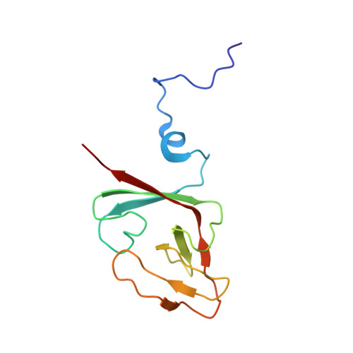
    1I4V

  • PubMed Abstract: 

    During the SOS response of Escherichia coli to DNA damage, the umuDC operon is induced, producing the trimeric protein complexes UmuD2C, a DNA damage checkpoint effector, and UmuD'2C (DNA polymerase V), which carries out translesion synthesis, the basis of 'SOS mutagenesis'. UmuD'2, the homodimeric component of DNA pol V, is produced from UmuD by RecA-facilitated self-cleavage, which removes the 24 N-terminal residues of UmuD. We report the solution structure of UmuD'2 (PDB ID 1I4V) and interactions within UmuD'-UmuD, a heterodimer inactive in translesion synthesis. The overall shape of UmuD'2 in solution differs substantially from the previously reported crystal structure, even though the topologies of the two structures are quite similar. Most significantly, the active site residues S60 and K97 do not point directly at one another in solution as they do in the crystal, suggesting that self-cleavage of UmuD might require RecA to assemble the active site. Structural differences between UmuD'2 and UmuD'- UmuD suggest that UmuD'2C and UmuD2C might achieve their different biological activities through distinct interactions with RecA and DNA pol III.


  • Organizational Affiliation
    • Department of Biological Chemistry and Molecular Pharmacology, Harvard Medical School, Boston, MA 02115, USA.

Macromolecule Content 

  • Total Structure Weight: 24.62 kDa 
  • Atom Count: 1,730 
  • Modeled Residue Count: 230 
  • Deposited Residue Count: 230 
  • Unique protein chains: 1

Macromolecules

Find similar proteins by:|  3D Structure
Entity ID: 1
MoleculeChains  Sequence LengthOrganismDetailsImage
UMUD' PROTEIN
A, B
115Escherichia coliMutation(s): 1 
Gene Names: UMUD
EC: 3.4.21
UniProt
Find proteins for P0AG11 (Escherichia coli (strain K12))
Explore P0AG11 
Go to UniProtKB:  P0AG11
Entity Groups
Sequence Clusters30% Identity50% Identity70% Identity90% Identity95% Identity100% Identity
UniProt GroupP0AG11
Sequence Annotations
Expand
Reference Sequence

Experimental Data & Validation

Experimental Data

  • Method: SOLUTION NMR
  • Conformers Calculated: 100 
  • Conformers Submitted: 20 
  • Selection Criteria: structures with the least restraint violations,structures with the lowest energy 

Structure Validation

View Full Validation Report


Entry History 

Deposition Data

Revision History  (Full details and data files)

  • Version 1.0: 2001-08-22
    Type: Initial release
  • Version 1.1: 2008-04-27
    Changes: Version format compliance
  • Version 1.2: 2011-07-13
    Changes: Version format compliance
  • Version 1.3: 2021-10-27
    Changes: Data collection, Database references, Derived calculations
  • Version 1.4: 2024-05-22
    Changes: Data collection