1HZM

STRUCTURE OF ERK2 BINDING DOMAIN OF MAPK PHOSPHATASE MKP-3: STRUCTURAL INSIGHTS INTO MKP-3 ACTIVATION BY ERK2


Experimental Data Snapshot

  • Method: SOLUTION NMR
  • Conformers Submitted: 

wwPDB Validation   3D Report Full Report

Currently 1HZM does not have a validation slider image.


This is version 1.3 of the entry. See complete history


Literature

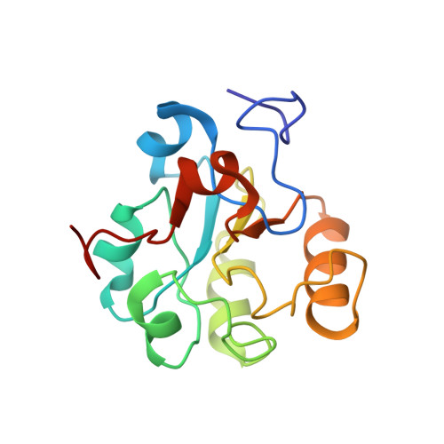
Solution structure of ERK2 binding domain of MAPK phosphatase MKP-3: structural insights into MKP-3 activation by ERK2.

Farooq, A.Chaturvedi, G.Mujtaba, S.Plotnikova, O.Zeng, L.Dhalluin, C.Ashton, R.Zhou, M.M.

(2001) Mol Cell 7: 387-399

  • DOI: https://doi.org/10.1016/s1097-2765(01)00186-1
  • Primary Citation of Related Structures:  
    1HZM

  • PubMed Abstract: 

    MAP kinases (MAPKs), which control mitogenic signal transduction in all eukaryotic organisms, are inactivated by dual specificity MAPK phosphatases (MKPs). MKP-3, a prototypical MKP, achieves substrate specificity through its N-terminal domain binding to the MAPK ERK2, resulting in the activation of its C-terminal phosphatase domain. The solution structure and biochemical analysis of the ERK2 binding (EB) domain of MKP-3 show that regions that are essential for ERK2 binding partly overlap with its sites that interact with the C-terminal catalytic domain, and that these interactions are functionally coupled to the active site residues of MKP-3. Our findings suggest a novel mechanism by which the EB domain binding to ERK2 is transduced to cause a conformational change of the C-terminal catalytic domain, resulting in the enzymatic activation of MKP-3.


  • Organizational Affiliation

    Structural Biology Program, Department of Physiology and Biophysics, Mount Sinai School of Medicine, New York University, New York, NY 10029, USA.


Macromolecules
Find similar proteins by:  (by identity cutoff)  |  3D Structure
Entity ID: 1
MoleculeChains Sequence LengthOrganismDetailsImage
DUAL SPECIFICITY PROTEIN PHOSPHATASE 6154Homo sapiensMutation(s): 0 
EC: 3.1.3.48 (PDB Primary Data), 3.1.3.16 (PDB Primary Data)
UniProt & NIH Common Fund Data Resources
Find proteins for Q16828 (Homo sapiens)
Explore Q16828 
Go to UniProtKB:  Q16828
PHAROS:  Q16828
GTEx:  ENSG00000139318 
Entity Groups  
Sequence Clusters30% Identity50% Identity70% Identity90% Identity95% Identity100% Identity
UniProt GroupQ16828
Sequence Annotations
Expand
  • Reference Sequence
Experimental Data & Validation

Experimental Data

  • Method: SOLUTION NMR
  • Conformers Submitted: 

Structure Validation

View Full Validation Report

Currently 1HZM does not have a validation slider image.


Entry History 

Deposition Data

Revision History  (Full details and data files)

  • Version 1.0: 2002-01-25
    Type: Initial release
  • Version 1.1: 2008-04-27
    Changes: Version format compliance
  • Version 1.2: 2011-07-13
    Changes: Version format compliance
  • Version 1.3: 2022-02-23
    Changes: Database references, Derived calculations