Role of charged residues at the OmpF porin channel constriction probed by mutagenesis and simulation.
Phale, P.S., Philippsen, A., Widmer, C., Phale, V.P., Rosenbusch, J.P., Schirmer, T.(2001) Biochemistry 40: 6319-6325
- PubMed: 11371193 Search on PubMed
- DOI: https://doi.org/10.1021/bi010046k
- Primary Citation Related Structures:
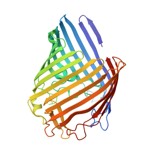
1HXT, 1HXU, 1HXX - PubMed Abstract:
The channel constriction of OmpF porin, a pore protein in the bacterial outer membrane, is highly charged due to the presence of three arginines (R42, R82, and R132) and two acidic residues (D113 and E117). The influence of these charges on ion conductance, ion selectivity, and voltage gating has been studied with mutants D113N/E117Q, R42A/R82A/R132A/D113N/E117Q, and V18K/G131K, which were designed to remove or add protein charge at the channel constriction. The crystal structures revealed no or only local changes compared to wild-type OmpF, thus allowing a comparative study. The single-channel conductance of the isosteric D113N/E117Q variant was found to be 2-fold reduced, and that of the pentuple mutant was 70% of the wild-type value, despite a considerably larger pore cross section. Ion selectivity was drastically altered by the mutations with cation/anion permeability ratios ranging from 1 to 12. Ion flow through these and eight other mutants, which have been characterized previously, was simulated by Brownian dynamics based on the detailed crystal structures. The calculated ion selectivity and relative channel conductance values agree well with the experimental data. This demonstrates that ion translocation through porin is mainly governed by pore geometry and charge, the two factors that are properly represented in the simulations.
- Division of Microbiology, Biozentrum, University of Basel, Klingelbergstrasse 70, CH-4056 Basel, Switzerland.
Organizational Affiliation: