1HTB

CRYSTALLIZATION OF HUMAN BETA3 ALCOHOL DEHYDROGENASE (10 MG/ML) IN 100 MM SODIUM PHOSPHATE (PH 7.5), 7.5 MM NAD+ AND 1 MM 4-IODOPYRAZOLE AT 25 C


Experimental Data Snapshot

  • Method: X-RAY DIFFRACTION
  • Resolution: 2.40 Å
  • R-Value Free: 0.243 
  • R-Value Work: 0.175 
  • R-Value Observed: 0.175 

wwPDB Validation   3D Report Full Report


Ligand Structure Quality Assessment 


This is version 1.3 of the entry. See complete history


Literature

X-ray structure of human beta3beta3 alcohol dehydrogenase. The contribution of ionic interactions to coenzyme binding.

Davis, G.J.Bosron, W.F.Stone, C.L.Owusu-Dekyi, K.Hurley, T.D.

(1996) J Biol Chem 271: 17057-17061

  • DOI: https://doi.org/10.1074/jbc.271.29.17057
  • Primary Citation of Related Structures:  
    1DEH, 1HTB

  • PubMed Abstract: 

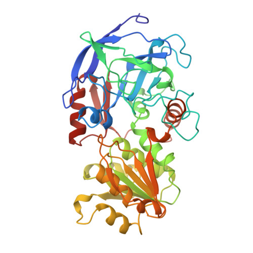
    The three-dimensional structure of the human beta3beta3 dimeric alcohol dehydrogenase (beta3) was determined to 2.4-A resolution. beta3 was crystallized as a ternary complex with the coenzyme NAD+ and the competitive inhibitor 4-iodopyrazole. beta3 is a polymorphic variant at ADH2 that differs from beta1 by a single amino acid substitution of Arg-369 --> Cys. The available x-ray structures of mammalian alcohol dehydrogenases show that the side chain of Arg-369 forms an ion pair with the NAD(H) pyrophosphate to stabilize the E.NAD(H) complex. The Cys-369 side chain of beta3 cannot form this interaction. The three-dimensional structures of beta3 and beta1 are virtually identical, with the exception that Cys-369 and two water molecules in beta3 occupy the position of Arg-369 in beta1. The two waters occupy the same positions as two guanidino nitrogens of Arg-369. Hence, the number of hydrogen bonding interactions between the enzyme and NAD(H) are the same for both isoenzymes. However, beta3 differs from beta1 by the loss of the electrostatic interaction between the NAD(H) pyrophosphate and the Arg-369 side chain. The equilibrium dissociation constants of beta3 for NAD+ and NADH are 350-fold and 4000-fold higher, respectively, than those for beta1. These changes correspond to binding free energy differences of 3.5 kcal/mol for NAD+ and 4.9 kcal/mol for NADH. Thus, the Arg-369 --> Cys substitution of beta3 isoenzyme destabilizes the interaction between coenzyme and beta3 alcohol dehydrogenase.


  • Organizational Affiliation

    Department of Biochemistry, Indiana University School of Medicine, Indianapolis, Indiana 46202-5122, USA.


Macromolecules
Find similar proteins by:  (by identity cutoff)  |  3D Structure
Entity ID: 1
MoleculeChains Sequence LengthOrganismDetailsImage
BETA3 ALCOHOL DEHYDROGENASE
A, B
374Homo sapiensMutation(s): 0 
Gene Names: HUMAN BETA3 CDNA
EC: 1.1.1.1
UniProt & NIH Common Fund Data Resources
Find proteins for P00325 (Homo sapiens)
Explore P00325 
Go to UniProtKB:  P00325
PHAROS:  P00325
GTEx:  ENSG00000196616 
Entity Groups  
Sequence Clusters30% Identity50% Identity70% Identity90% Identity95% Identity100% Identity
UniProt GroupP00325
Sequence Annotations
Expand
  • Reference Sequence
Small Molecules
Ligands 4 Unique
IDChains Name / Formula / InChI Key2D Diagram3D Interactions
NAD
Query on NAD

Download Ideal Coordinates CCD File 
E [auth A],
J [auth B]
NICOTINAMIDE-ADENINE-DINUCLEOTIDE
C21 H27 N7 O14 P2
BAWFJGJZGIEFAR-NNYOXOHSSA-N
PYZ
Query on PYZ

Download Ideal Coordinates CCD File 
F [auth A],
K [auth B]
4-IODOPYRAZOLE
C3 H3 I N2
LLNQWPTUJJYTTE-UHFFFAOYSA-N
ZN
Query on ZN

Download Ideal Coordinates CCD File 
C [auth A],
D [auth A],
G [auth B],
H [auth B]
ZINC ION
Zn
PTFCDOFLOPIGGS-UHFFFAOYSA-N
CL
Query on CL

Download Ideal Coordinates CCD File 
I [auth B]CHLORIDE ION
Cl
VEXZGXHMUGYJMC-UHFFFAOYSA-M
Binding Affinity Annotations 
IDSourceBinding Affinity
NAD Binding MOAD:  1HTB Kd: 6.70e+5 (nM) from 1 assay(s)
Experimental Data & Validation

Experimental Data

  • Method: X-RAY DIFFRACTION
  • Resolution: 2.40 Å
  • R-Value Free: 0.243 
  • R-Value Work: 0.175 
  • R-Value Observed: 0.175 
  • Space Group: P 1
Unit Cell:
Length ( Å )Angle ( ˚ )
a = 54.03α = 92.71
b = 44.43β = 103.17
c = 92.74γ = 69.15
Software Package:
Software NamePurpose
RAXISdata collection
RIGAKUdata collection
JAPANdata collection
X-PLORmodel building
X-PLORrefinement
R-AXISdata reduction
X-PLORphasing

Structure Validation

View Full Validation Report


Ligand Structure Quality Assessment 


Entry History 

Deposition Data

Revision History  (Full details and data files)

  • Version 1.0: 1995-12-07
    Type: Initial release
  • Version 1.1: 2008-03-03
    Changes: Version format compliance
  • Version 1.2: 2011-07-13
    Changes: Version format compliance
  • Version 1.3: 2024-02-07
    Changes: Data collection, Database references, Derived calculations, Other