1HST | pdb_00001hst

CRYSTAL STRUCTURE OF GLOBULAR DOMAIN OF HISTONE H5 AND ITS IMPLICATIONS FOR NUCLEOSOME BINDING


Experimental Data Snapshot

  • Method: X-RAY DIFFRACTION
  • Resolution: 2.60 Å
  • R-Value Work: 
    0.270 (Depositor) 
  • R-Value Observed: 
    0.270 (Depositor) 

wwPDB Validation 3D Report Full Report

Validation slider image for 1HST

This is version 1.3 of the entry. See complete history

Literature

Crystal structure of globular domain of histone H5 and its implications for nucleosome binding.

Ramakrishnan, V.Finch, J.T.Graziano, V.Lee, P.L.Sweet, R.M.

(1993) Nature 362: 219-223

  • DOI: https://doi.org/10.1038/362219a0
  • Primary Citation Related Structures: 
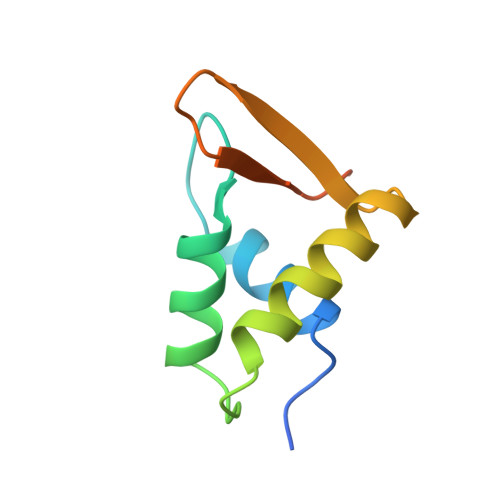
    1HST

  • PubMed Abstract: 

    The structure of GH5, the globular domain of the linker histone H5, has been solved to 2.5 A resolution by multiwavelength anomalous diffraction on crystals of the selenomethionyl protein. The structure shows a striking similarity to the DNA-binding domain of the catabolite gene activator protein CAP, thereby providing a possible model for the binding of GH5 to DNA.


  • Organizational Affiliation
    • Biology Department, Brookhaven National Laboratory, Upton, New York 11973.

Macromolecule Content 

  • Total Structure Weight: 19.58 kDa 
  • Atom Count: 1,128 
  • Modeled Residue Count: 148 
  • Deposited Residue Count: 180 
  • Unique protein chains: 1

Macromolecules

Find similar proteins by:|  3D Structure
Entity ID: 1
MoleculeChains  Sequence LengthOrganismDetailsImage
HISTONE H5
A, B
90Gallus gallusMutation(s): 0 
UniProt
Find proteins for P02259 (Gallus gallus)
Explore P02259 
Go to UniProtKB:  P02259
Entity Groups
Sequence Clusters30% Identity50% Identity70% Identity90% Identity95% Identity100% Identity
UniProt GroupP02259
Sequence Annotations
Expand
Reference Sequence

Experimental Data & Validation

Experimental Data

  • Method: X-RAY DIFFRACTION
  • Resolution: 2.60 Å
  • R-Value Work:  0.270 (Depositor) 
  • R-Value Observed: 0.270 (Depositor) 
Space Group: P 21 21 2
Unit Cell:
Length ( Å )Angle ( ˚ )
a = 68.5α = 90
b = 80.4β = 90
c = 38.9γ = 90
Software Package:
Software NamePurpose
X-PLORmodel building
X-PLORrefinement
X-PLORphasing

Structure Validation

View Full Validation Report


Entry History 

Deposition Data

Revision History  (Full details and data files)

  • Version 1.0: 1993-10-31
    Type: Initial release
  • Version 1.1: 2008-03-24
    Changes: Version format compliance
  • Version 1.2: 2011-07-13
    Changes: Version format compliance
  • Version 1.3: 2024-02-07
    Changes: Data collection, Database references, Other