1HCQ

THE CRYSTAL STRUCTURE OF THE ESTROGEN RECEPTOR DNA-BINDING DOMAIN BOUND TO DNA: HOW RECEPTORS DISCRIMINATE BETWEEN THEIR RESPONSE ELEMENTS


Experimental Data Snapshot

  • Method: X-RAY DIFFRACTION
  • Resolution: 2.40 Å
  • R-Value Work: 0.204 
  • R-Value Observed: 0.204 

wwPDB Validation   3D Report Full Report


This is version 1.3 of the entry. See complete history


Literature

The crystal structure of the estrogen receptor DNA-binding domain bound to DNA: how receptors discriminate between their response elements.

Schwabe, J.W.Chapman, L.Finch, J.T.Rhodes, D.

(1993) Cell 75: 567-578

  • DOI: https://doi.org/10.1016/0092-8674(93)90390-c
  • Primary Citation of Related Structures:  
    1HCQ

  • PubMed Abstract: 

    The nuclear hormone receptors are a superfamily of ligand-activated DNA-binding transcription factors. We have determined the crystal structure (at 2.4 A) of the fully specific complex between the DNA-binding domain from the estrogen receptor and DNA. The protein binds as a symmetrical dimer to its palindromic binding site consisting of two 6 bp consensus half sites with three intervening base pairs. This structure reveals how the protein recognizes its own half site sequence rather than that of the related glucocorticoid receptor, which differs by only two base pairs. Since all nuclear hormone receptors recognize one or the other of these two consensus half site sequences, this recognition mechanism applies generally to the whole receptor family.


  • Organizational Affiliation

    Medical Research Council Laboratory of Molecular Biology, Cambridge, England.


Macromolecules

Find similar proteins by:  (by identity cutoff)  |  3D Structure
Entity ID: 3
MoleculeChains Sequence LengthOrganismDetailsImage
PROTEIN (ESTROGEN RECEPTOR)E [auth A],
F [auth B],
G [auth E],
H [auth F]
84Homo sapiensMutation(s): 0 
UniProt & NIH Common Fund Data Resources
Find proteins for P03372 (Homo sapiens)
Explore P03372 
Go to UniProtKB:  P03372
PHAROS:  P03372
GTEx:  ENSG00000091831 
Entity Groups  
Sequence Clusters30% Identity50% Identity70% Identity90% Identity95% Identity100% Identity
UniProt GroupP03372
Sequence Annotations
Expand
  • Reference Sequence

Find similar nucleic acids by:  Sequence   |   3D Structure  

Entity ID: 1
MoleculeChains LengthOrganismImage
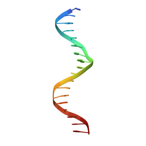
DNA (5'-D(*CP*CP*AP*GP*GP*TP*CP*AP*CP*AP*GP*TP*GP*AP*CP*CP*T P*G)-3')A [auth C],
C [auth G]
18N/A
Sequence Annotations
Expand
  • Reference Sequence

Find similar nucleic acids by:  Sequence   |   3D Structure  

Entity ID: 2
MoleculeChains LengthOrganismImage
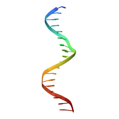
DNA (5'-D(*CP*CP*AP*GP*GP*TP*CP*AP*CP*TP*GP*TP*GP*AP*CP*CP*T P*G)-3')B [auth D],
D [auth H]
18N/A
Sequence Annotations
Expand
  • Reference Sequence
Experimental Data & Validation

Experimental Data

  • Method: X-RAY DIFFRACTION
  • Resolution: 2.40 Å
  • R-Value Work: 0.204 
  • R-Value Observed: 0.204 
  • Space Group: P 21 21 21
Unit Cell:
Length ( Å )Angle ( ˚ )
a = 51.6α = 90
b = 90.8β = 90
c = 114.6γ = 90
Software Package:
Software NamePurpose
X-PLORrefinement
TNTrefinement

Structure Validation

View Full Validation Report


Entry History 

Deposition Data

Revision History  (Full details and data files)

  • Version 1.0: 1995-11-23
    Type: Initial release
  • Version 1.1: 2008-05-22
    Changes: Version format compliance
  • Version 1.2: 2011-07-13
    Changes: Version format compliance
  • Version 1.3: 2022-12-21
    Changes: Database references, Derived calculations