1H8U | pdb_00001h8u

Crystal Structure of the Eosinophil Major Basic Protein at 1.8A: An Atypical Lectin with a Paradigm Shift in Specificity


Experimental Data Snapshot

  • Method: X-RAY DIFFRACTION
  • Resolution: 1.80 Å
  • R-Value Free: 
    0.264 (Depositor) 
  • R-Value Work: 
    0.234 (Depositor) 
  • R-Value Observed: 
    0.234 (Depositor) 

Starting Model: experimental
View more details

wwPDB Validation 3D Report Full Report

Validation slider image for 1H8U

This is version 1.4 of the entry. See complete history

Literature

Crystal Structure of the Eosinophil Major Basic Protein at 1.8A. An Atypical Lectin with a Paradigm Shift in Specificity

Swaminathan, G.J.Weaver, A.J.Loegering, D.A.Checkel, J.L.Leonidas, D.D.Gleich, G.J.Acharya, K.R.

(2001) J Biological Chem 276: 26197

  • DOI: https://doi.org/10.1074/jbc.M100848200
  • Primary Citation Related Structures: 
    1H8U

  • PubMed Abstract: 

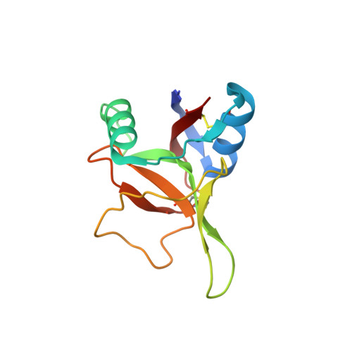
    The eosinophil major basic protein (EMBP) is the predominant constituent of the crystalline core of the eosinophil primary granule. EMBP is directly implicated in epithelial cell damage, exfoliation, and bronchospasm in allergic diseases such as asthma. Here we report the crystal structure of EMBP at 1.8 A resolution, and show that it is similar to that of members of the C-type lectin superfamily with which it shares minimal amino acid sequence identity (approximately 15--28%). However, this protein lacks a Ca(2+)/carbohydrate-binding site. Our analysis suggests that EMBP specifically binds heparin. Based on our results, we propose a possible new function for this protein, which is likely to have implications for EMBP function.


  • Organizational Affiliation
    • Department of Biology and Biochemistry, University of Bath, Claverton Down, Bath BA2 7AY, United Kingdom.

Macromolecule Content 

  • Total Structure Weight: 28.59 kDa 
  • Atom Count: 2,124 
  • Modeled Residue Count: 231 
  • Deposited Residue Count: 234 
  • Unique protein chains: 1

Macromolecules

Find similar proteins by:|  3D Structure
Entity ID: 1
MoleculeChains  Sequence LengthOrganismDetailsImage
EOSINOPHIL GRANULE MAJOR BASIC PROTEIN 1
A, B
117Homo sapiensMutation(s): 0 
UniProt & NIH Common Fund Data Resources
Find proteins for P13727 (Homo sapiens)
Explore P13727 
Go to UniProtKB:  P13727
PHAROS:  P13727
GTEx:  ENSG00000186652 
Entity Groups
Sequence Clusters30% Identity50% Identity70% Identity90% Identity95% Identity100% Identity
UniProt GroupP13727
Sequence Annotations
Expand
Reference Sequence

Small Molecules

Ligands 2 Unique
IDChains Name / Formula / InChI Key2D Diagram3D Interactions
SO4

Query on SO4


Download:Ideal Coordinates CCD File
C [auth A]
D [auth A]
E [auth A]
F [auth A]
G [auth A]
C [auth A],
D [auth A],
E [auth A],
F [auth A],
G [auth A],
J [auth B],
K [auth B],
L [auth B]
SULFATE ION
O4 S
QAOWNCQODCNURD-UHFFFAOYSA-L
GOL

Query on GOL


Download:Ideal Coordinates CCD File
H [auth A],
I [auth A]
GLYCEROL
C3 H8 O3
PEDCQBHIVMGVHV-UHFFFAOYSA-N

Experimental Data & Validation

Experimental Data

  • Method: X-RAY DIFFRACTION
  • Resolution: 1.80 Å
  • R-Value Free:  0.264 (Depositor) 
  • R-Value Work:  0.234 (Depositor) 
  • R-Value Observed: 0.234 (Depositor) 
Space Group: C 1 2 1
Unit Cell:
Length ( Å )Angle ( ˚ )
a = 74.328α = 90
b = 57.487β = 113.2
c = 60.961γ = 90
Software Package:
Software NamePurpose
CNSrefinement
DENZOdata reduction
SCALEPACKdata scaling
AMoREphasing

Structure Validation

View Full Validation Report


Entry History 

Deposition Data

Revision History  (Full details and data files)

  • Version 1.0: 2001-07-17
    Type: Initial release
  • Version 1.1: 2011-05-07
    Changes: Version format compliance
  • Version 1.2: 2011-07-13
    Changes: Version format compliance
  • Version 1.3: 2023-12-13
    Changes: Data collection, Database references, Refinement description
  • Version 1.4: 2024-10-23
    Changes: Structure summary